Figure 4. Representative electron microscopic images of autophagic vacuoles in murine cytomegalovirus-infected and -uninfected retinal
pigment epithelial (RPE) cells. RPE cells were cultured in normal medium or infected with murine cytomegalovirus (MCMV) at
low multiplicity of infection (MOI) = 1 for 3 days and then fixed and processed for electron microscopy. A: Representative view of normal RPE cells. B: Higher magnification view of autophagic vacuoles in MCMV-infected cells. Panel b is the enlarged view of the rectangle in
panel a. Panel c is the enlarged view of the rectangle in panel a. Black arrow in panel b: autophagic vacuole; black arrows
in panel c: viral particles; Nu: nucleus; black arrow in panel d: phagosome. C: Higher magnification view of autophagic vacuoles containing viral particle. White arrow: viral particle. D: Quantification of autophagic vacuoles for a minimum of 20 MCMV-infected or uninfected cells. The number of autophagic vacuoles
per cell was determined in electron micrographs. ***p<0.001, two-tailed Student t test. Data are shown as mean±SEM (n=3).
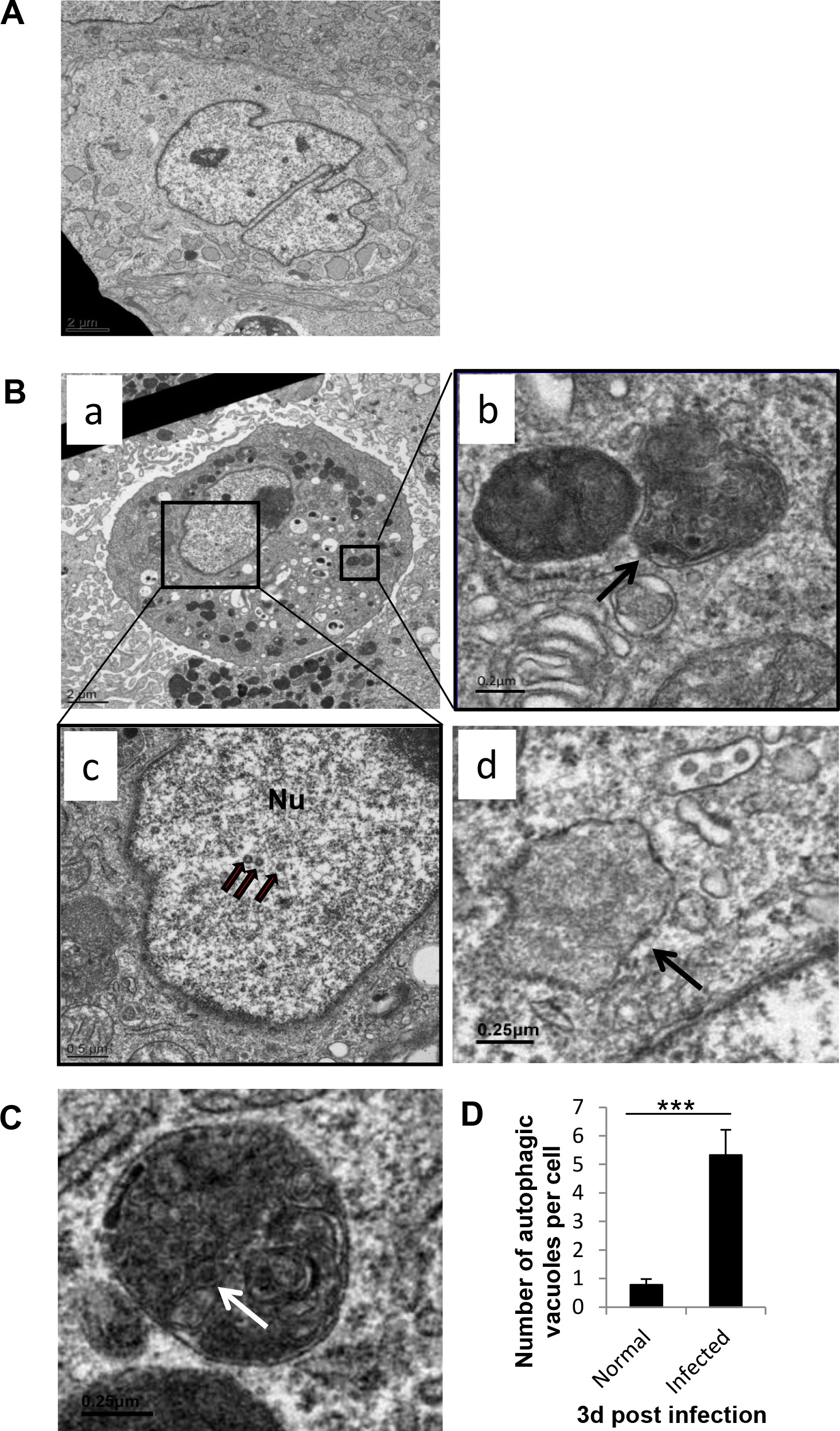
 Figure 4 of
Mo, Mol Vis 2014; 20:1161-1173.
Figure 4 of
Mo, Mol Vis 2014; 20:1161-1173.