Figure 3. Representative images of retinal pigment epithelial (RPE) cells with green fluorescent protein-light chain 3 (GFP-LC3) puncta.
RPE cells cultured in normal medium were transiently transfected with green fluorescent protein–light chain 3 (GFP-LC3) plasmid
(CT) or infected with murine cytomegalovirus (MCMV) at low multiplicity of infection (MOI) = 1 for 24 h. A: Representative images (×630) of RPE cells with GFP-LC3 puncta. White circle: GFP-LC3 positive puncta. B: Quantification of autophagy in RPE cells transiently transfected with GFP-LC3 plasmid in normal medium or infected with
MCMV at MOI = 1 for 24 h. ***p<0.001, two-tailed Student t test. Data are shown as mean±SEM (n=3).
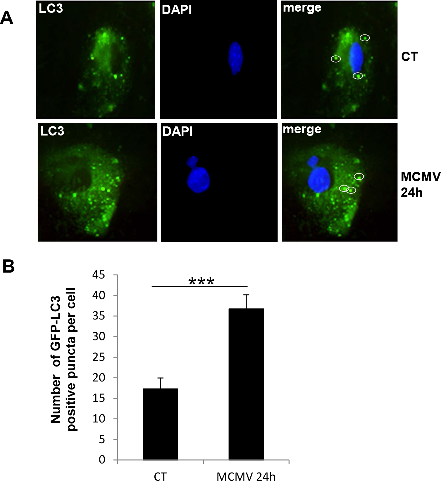
 Figure 3 of
Mo, Mol Vis 2014; 20:1161-1173.
Figure 3 of
Mo, Mol Vis 2014; 20:1161-1173.