Figure 2. Autophagic flux during murine cytomegalovirus infection of retinal pigment epithelial (RPE) cells. RPE cells were infected
with murine cytomegalovirus (MCMV) at low multiplicity of infection (MOI) = 1 in normal medium or in medium containing chloroquine
(CQ, 10−6 M) or ammonium chloride (NH4Cl, 10−6 M) to block autophagic flux for 6 h (A), 12 h (B), 24 h (C), 2 days (D), and 3 days (E). Expression of processed light-chain 3B (LC3B) was monitored.
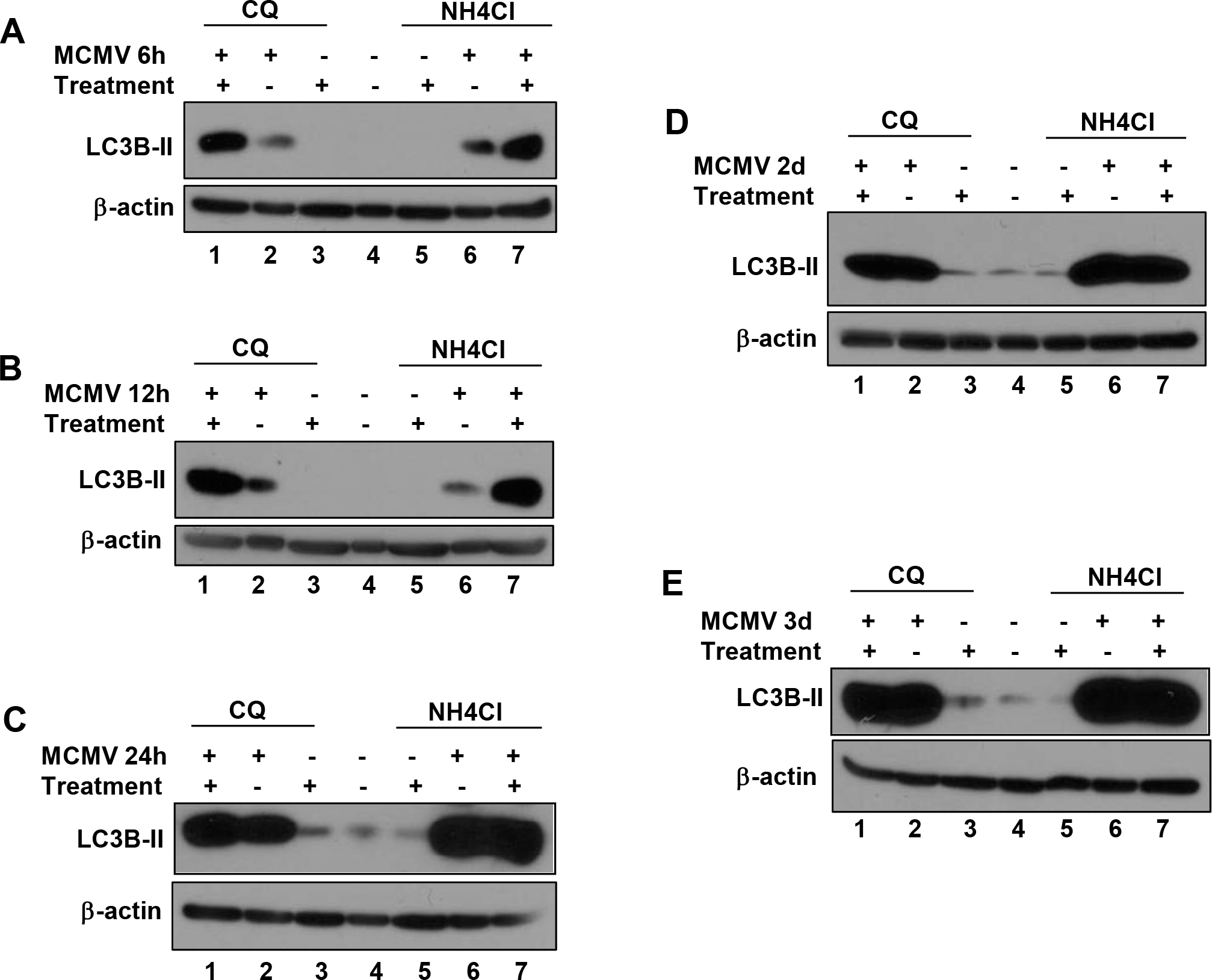
 Figure 2 of
Mo, Mol Vis 2014; 20:1161-1173.
Figure 2 of
Mo, Mol Vis 2014; 20:1161-1173.