Figure 1. Autophagic response to different stimuli. A: Retinal pigment epithelial (RPE) cells were cultured in normal medium (CT) and treated with chloroquine (CQ, 10−6 M) or serum deprivation (SD) for 24 h. Expression of processed light-chain 3B (LC3B) was monitored. B: Semi-quantitative analysis of western blot for LC3B protein expression in the RPE cells from the control, CQ-treated, and
SD-treated groups. C: Representative images (×200) of murine cytomegalovirus (MCMV)-infected RPE cells. RPE cells were infected with MCMV at multiplicity
of infection (MOI) = 1 for 1, 2, 3, and 4 days. Black arrow: infected cell. 1d: 1 day postinfection; 2d: 2 days postinfection;
3d: 3 days postinfection; 4d: 4 days postinfection. *p<0.05, ***p<0.001, ANOVA. Data are shown as mean±SEM (n=3).
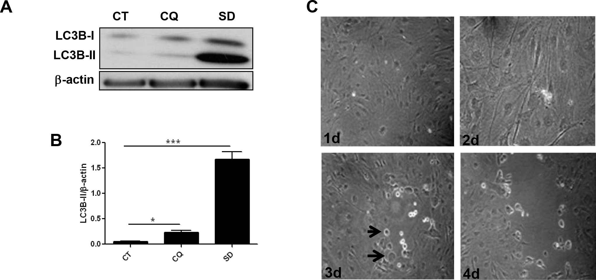
 Figure 1 of
Mo, Mol Vis 2014; 20:1161-1173.
Figure 1 of
Mo, Mol Vis 2014; 20:1161-1173.