Figure 6. Indirect immunofluorescence evaluation of apelin and fibronectin distribution in human epiretinal membranes (ERMs) derived
from patients with proliferative diabetic retinopathy (PDR). Cryosections were double-probed with antibodies against (A) apelin and (B) fibronectin. Nuclei were detected using 4', 6-diamidino-2-phenylindole (DAPI). C: Merged images contain three color channels representing apelin (red), fibronectin (green), and DAPI (blue). The arrow showed
apelin was not co-expressed with fibronectin in ERMs from PDR patients. Scale bar represents 100 μm.
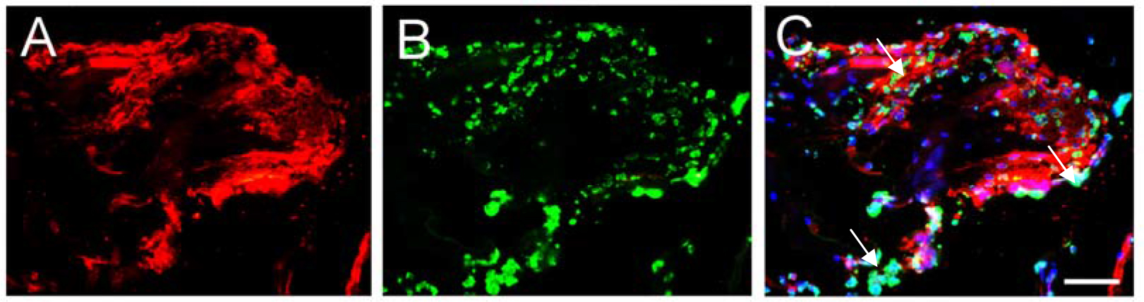
 Figure 6 of
Lu, Mol Vis 2014; 20:1122-1131.
Figure 6 of
Lu, Mol Vis 2014; 20:1122-1131.