Figure 5. Fluorescence immunostaining of human epiretinal membranes (ERMs) in negative controls. No immunostaining was observed in negative
controls (arrow), which were performed by substituting the primary antiserum with phosphate buffered saline. Secondary antibodies
were used cyanogen (CY) 3-conjugated goat anti-rabbit-fluorescein isothiocyanate (A) and FITC-conjugated goat anti-mouse-tetramethyl rhodamine isothiocyanate (B). Nuclei were detected using DAPI. Merged images (C), DAPI (blue). Scale bar represents 100 μm.
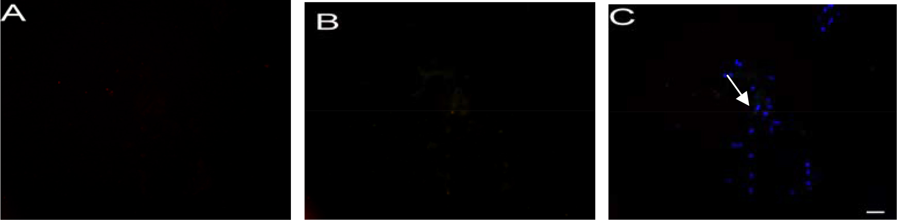
 Figure 5 of
Lu, Mol Vis 2014; 20:1122-1131.
Figure 5 of
Lu, Mol Vis 2014; 20:1122-1131.