Figure 4. Indirect immunofluorescence evaluation of apelin and glial fibrillary acidic protein distribution in human epiretinal membranes
(ERMs) derived from patients with proliferative diabetic retinopathy (PDR; A, B, C). Apelin and glial fibrillary acidic protein (GFAP) distribution in idiopathic epiretinal membranes (ERMs) from the control
subjects (D, E, F). ERMs from patients with proliferative diabetic retinopathy (PDR) after intravitreal injection of bevacizumab (G, H, I). Cryosections were double-probed with antibodies against apelin (A, D, G), GFAP (B), and GFAP (E, H), and were detected with fluorochrome-conjugated secondary antibodies. Nuclei were detected using 4', 6-diamidino-2-phenylindole
(DAPI) (C, F, I). The arrow in C showed apelin was co-expressed with GFAP. The arrow in F and I showed apelin was not expressed in idiopathic ERMs and ERMs from PDR patients after intravitreal injection of bevacizumab,
respectively. Scale bar represents 100 μm.
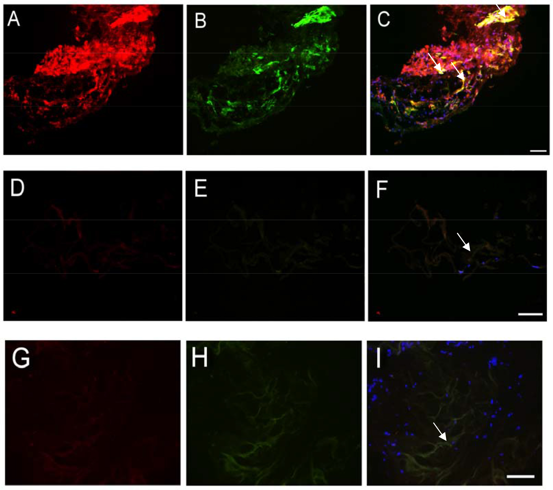
 Figure 4 of
Lu, Mol Vis 2014; 20:1122-1131.
Figure 4 of
Lu, Mol Vis 2014; 20:1122-1131.