Figure 3. Immunofluorescence staining for apelin, cytokeratin (CK), platelet endothelial cell adhesion molecule-1 (CD31), and vascular
endothelial growth factor (VEGF) in proliferative diabetic retinopathy (PDR) epiretinal membranes. Cryosections were double-probed
with antibodies against apelin (A, D, G) and (B) CK, (E) CD31, and (H) VEGF and detected using fluorochrome-conjugated secondary antibodies. Nuclei were labeled using 4', 6-diamidino-2-phenylindole
(DAPI). Merged images (C, F, I) contain three colour channels representing apelin (red), CK, CD31, and VEGF (green), and DAPI (blue). The arrow in C, F, I showed apelin was co-expressed with CK, CD31, and VEGF in in ERMs from patients with PDR, respectively. Scale bar represents
50 μm.
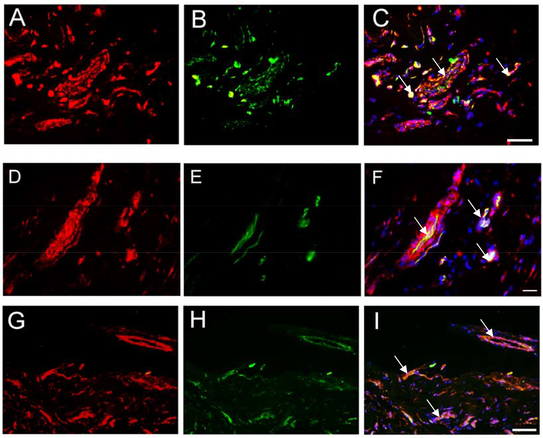
 Figure 3 of
Lu, Mol Vis 2014; 20:1122-1131.
Figure 3 of
Lu, Mol Vis 2014; 20:1122-1131.