Figure 2. Histopathologic findings in fibrovascular membranes of proliferative diabetic retinopathy (PDR; A, B) and in idiopathic epiretinal membranes (ERMs; C). A: H&E staining shows densely cellular tissue in ERMs from PDR patients (arrow). B: H&E staining shows highly vascularized tissue and large-calibre vessels and gliosis in ERMs from patients with PDR (arrow).
C: H&E staining shows sparse cellular tissue in idiopathic ERMs derived from the control subjects.
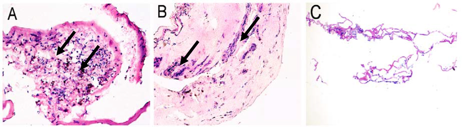
 Figure 2 of
Lu, Mol Vis 2014; 20:1122-1131.
Figure 2 of
Lu, Mol Vis 2014; 20:1122-1131.