Figure 3. Activation of extracellular signal-regulated kinases 1 and 2 (ERK1/2) signaling pathways in the retinas of STZ rats. A: Western blot analysis of the ERK1/2 phosphorylation in the retinas of diabetic rats from 3 days to 6 weeks after the induction
of diabetes. B: The levels of ERK1/2 phosphorylation were increased in the retinas of diabetic rats at 1 week and 2 weeks after the induction
of diabetes and displayed a time-dependent trend. Each column denotes the mean ± SD (n = 6). C: Immunofluorescence showed that ERK1/2 expression located in the retinal ganglion cell layer increased in 1 week diabetic
rats compared with control. Scale bar, 100 μm. D: Changes in ERK1/2 phosphorylation in the retinas of STZ rats transduced with LV.shScrambled or LV. shGPR91 using western
blotting. Rats were analyzed after 1 week after the induction of diabetes. E: The increases in p-ERK1/2 expression were significantly blocked by GPR91 shRNA in the 1 week diabetic rats. Each column
denotes the mean ± SD (n = 6). **p<0.01 versus control. ##p<0.01 versus LV.shScrambled group rats.
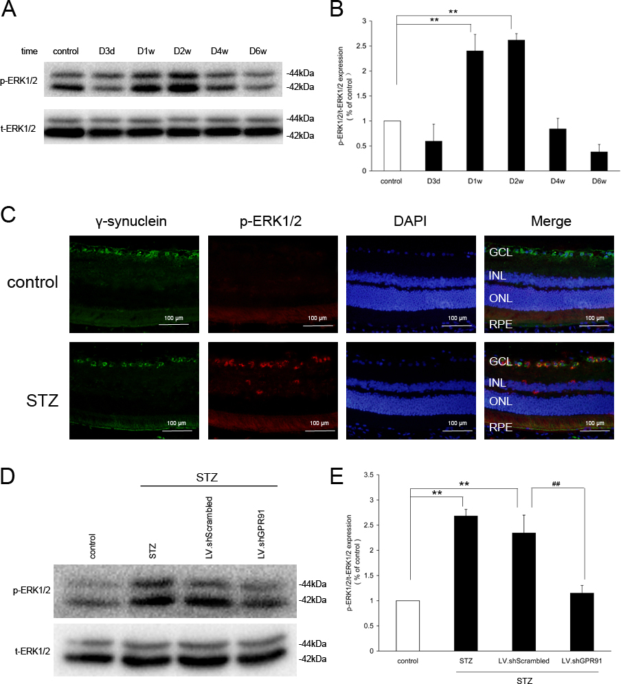
 Figure 3 of
Li, Mol Vis 2014; 20:1109-1121.
Figure 3 of
Li, Mol Vis 2014; 20:1109-1121.