Figure 2. Attenuation of retinal vascular dysfunction and inhibition of vascular endothelial growth factor (VEGF) expression in STZ-induced
diabetic rats following intravitreal injection of shRNA lentiviral particles targeting GPR91 (LV.shGPR91) particles. A: Light microscopy analysis of retinas from animals in each group. Compared with the non-diabetic rats, hematoxylin and eosin
(HE) staining revealed that the retina tissue developed telangiectatic vessels in the inner layer of retinas and the irregular arrangement
of cells in the inner nuclear layer in diabetic rats for the 14 week experiment. Treatment with GPR91 shRNA attenuated the
retinal vascular dysfunction compare to LV.shScrambled group rats but had no effect on the damage of the inner nuclear layer.
The black arrows indicate the telangiectatic vessel. Scale bar, 50 μm. B: The number of blood vessel profiles (BVPs) per unit area of the inner retina was increased in the 14 week diabetic rats
compared with the non-diabetic rats. Treatment with GPR91 shRNA reduced the number of BVPs per unit area of the inner retina
compared to rats in the LV.shScrambled group. Each column denotes the mean ± SD (n = 6). C: Transmission electron micrograph (TEM) images of the retinal microvasculature in samples from each group. TEM examinations
revealed that swelling was observed in the mitochondria of the pericytes and endothelial cells and that the mitochondrial
membrane was ruptured in the 14 week diabetic rats. Treatment with GPR91 shRNA significantly attenuated ultrastructural changes
of the retina compared to the LV.shScrambled group rats. The black arrow denotes the segment of the outer capillary basement membrane between the endothelial cells and glia limitans, which was used
to measure the basement membrane thickness. The white square frame denotes swelling in the mitochondria of the vascular pericytes and endothelial cells. Scale bar, 1 μm. D: In the 14 week diabetic retinal capillary, the basement membrane thickness (BMT) was significantly greater compared with
the non-diabetic rats. Treatment with GPR91 shRNA significantly decreased the BMT compared to the LV.shScrambled group rats.
Each column denotes the mean ± SD (n = 6). E: An increase in retinal vascular permeability was detected by Evans blue dye in the diabetic rats at the completion of the
experiment. Treatment with GPR91 shRNA significantly reduced Evans blue dye permeability compared with the scrambled shRNA
group. Each column denotes the mean ± SD (n = 6). F: qRT-PCR analysis for VEGF mRNA in the 4 week diabetic rat retinas was significantly upregulated compared with the non-diabetic
rats. The levels of VEGF mRNA in the 4 week STZ rats treated with GPR91 shRNA were reduced by approximately 25% compared with
the scrambled shRNA group. Each column denotes the mean ± SD (n = 6). G: Enzyme-linked immunosorbent assay analysis of vitreal VEGF release in vitreous. The retinal expression of VEGF protein was
increased in diabetic rats for the 4 week experiment compared with the non-diabetic rats. GPR91 knockdown significantly reduced
VEGF protein expression by approximately 75% compared with the scrambled shRNA group. Each column denotes the mean ± SD (n
= 6). **p<0.01 versus control. #p<0.05 versus LV.shScrambled group rats. ##p<0.01 versus LV.shScrambled group rats.
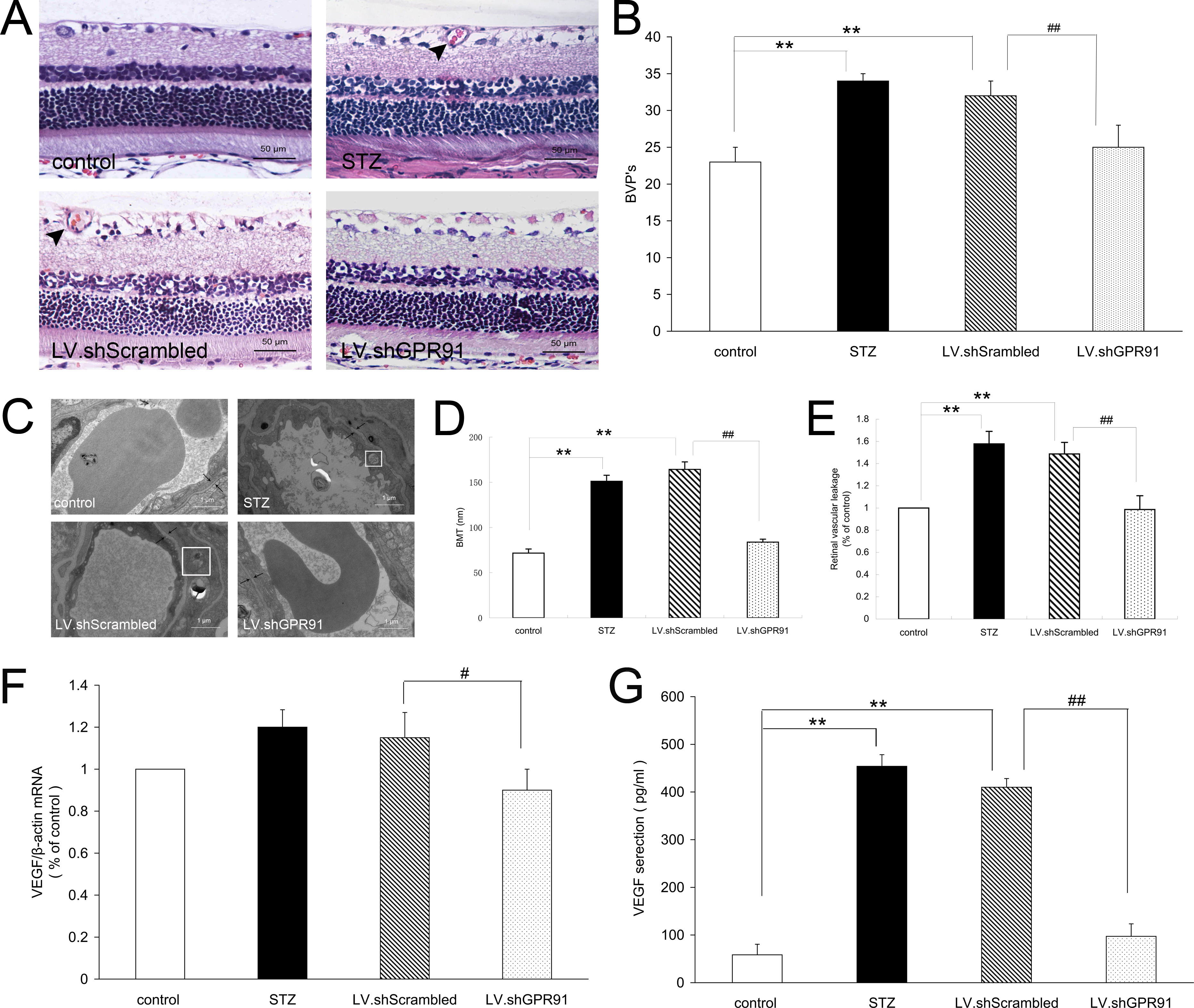
 Figure 2 of
Li, Mol Vis 2014; 20:1109-1121.
Figure 2 of
Li, Mol Vis 2014; 20:1109-1121.