Figure 8. Intravitreal injection of bone morphogenetic protein 7 (BMP7) into murine eyes leads to reactive gliosis. A: RNA from adult eyes injected with vehicle or BMP7 was isolated 3 and 7 days following injections and real time quantitative
PCR (RT-qPCR) was performed using the reactive gliosis panel described in previous figures. Graphs have been normalized to
β2 Microglobulin and all values are relative to vehicle-treated eyes from the same post-injection day. Each sample was run in triplicate and
experiment repeated 3 times for each gene. Values represented are means ±SEM. Unpaired t test was performed between the control and treated groups with * denoting a p value<0.05 and ** denoting a p value<0.005.
Any value above 1.0 represents an increase in mRNA levels, while a level below 1.0 represents a decrease. At 3 days following
injection, there was a significant decrease in paired-homeobox 2 (Pax2) and galectin 3 (Gal3). In contrast, by 7 days following injection there were significant increases in thioredoxin-interacting protein (Txnip), glutamate-aspartate transporter (Glast), glutamine synthetase (GS), matrix metalloproteinase 9 (Mmp9), lipocalin 2 (Lcn2), potassium inwardly rectifying channel 2.1 (Kir2.1), and matrix metalloproteinase 14 (Mmp14) and decreases in toll-like receptor 4 (Tlr4), Pax2, and epidermal growth factor receptor (Egfr). B: Immunolabel of retinas from 3 day and 7 day vehicle and BMP7 injected eyes for a subset of the gliosis markers. To confirm
some of the changes seen at the mRNA level, eyes injected with vehicle or BMP7 were fixed 3 or 7 days after injection and
immunolabeled for GFAP, S100β, or NEUROCAN (NCAN). At 3 days following injection, GFAP, S100β and NCAN immunolabel looks similar
in vehicle- and BMP7-injected eyes. In contrast, by 7 days, sections immunolabeled for GFAP, S100β, and NCAN showed an increase
in labeling in BMP7-injected eyes in comparison to vehicle-injected. n=3 different retinas for each label. Scale bar A=50 μm applies to all panels. C: western blot analysis was performed for GFAP, TXNIP, S100β, and PAX2, with β-TUBULIN used as a loading control. Densitometric
data shown are means±SEM of 3 trials. Unpaired t test was performed between the control and treated groups with * denoting a p value<0.05 and ** denoting a p value<0.005.
Densitometric analysis of the blots showed a statistical increase in protein levels of S100β at the 3 day stage. While there
did appear to be an increase in GFAP, S100β, and TXNIP at 7 days following injection in the BMP7-treated retinas as compared
to vehicle-injected, none of the changes in protein levels was statistically significant.
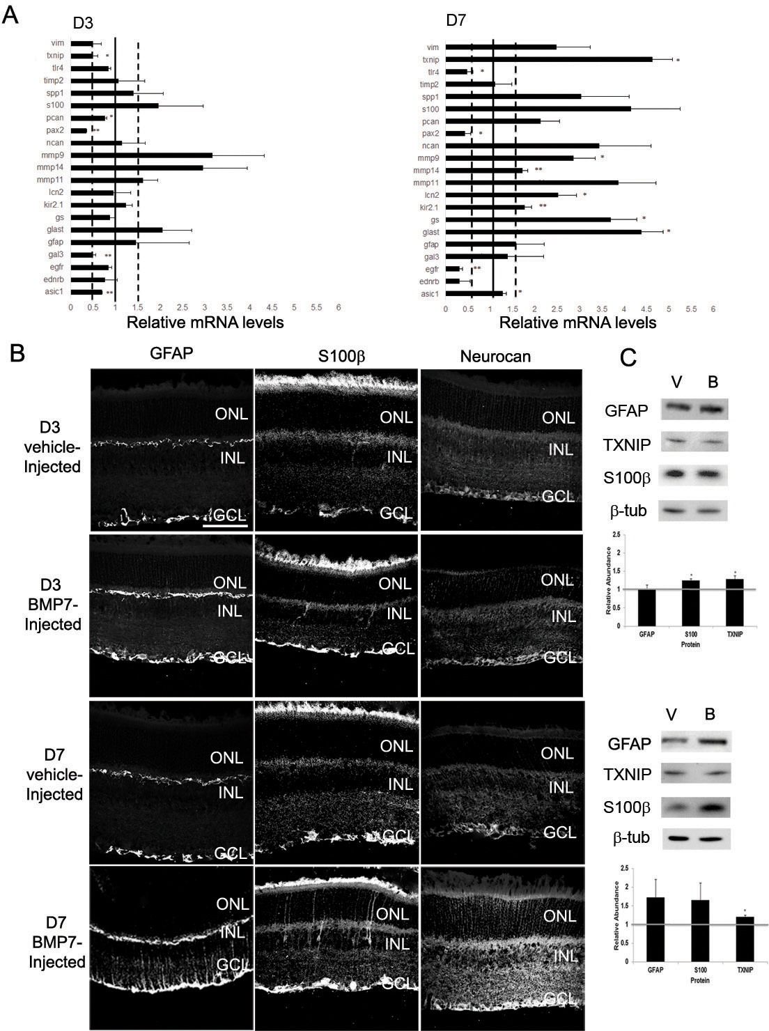
 Figure 8 of
Dharmarajan, Mol Vis 2014; 20:1085-1108.
Figure 8 of
Dharmarajan, Mol Vis 2014; 20:1085-1108.