Figure 6. Canonical bone morphogenetic protein (BMP) signaling is activated in the Müller glia in BMP7 injected murine eyes. Sections
of retina from adult mouse eyes injected with vehicle or BMP7 and harvested 3 or 7 days after injections were double-labeled
with antibodies against phospho-SMAD1/5/8 (green) and the nuclear marker sex determining region Y box 2 (SOX2; red) which
labels Müller glia, retinal astrocytes and cholinergic amacrine cells . While the vehicle-injected control showed phospho-SMAD
1/5/8 labeled cells primarily localized in the ganglion cell layer (GCL), with little or no co-label with the SOX2 (+) cells,
in the D3 and D7 BMP7-injected retinas, phospho-SMAD1/5/8 label was also detectable in the inner nuclear layer (arrows INL).
A sub population of the cells in the INL which were positive for phospho-SMAD1/5/8 were also SOX2 positive. n=3 different
retinas for each immunolabel. Scale Bar A=50 μm applies to all panels.
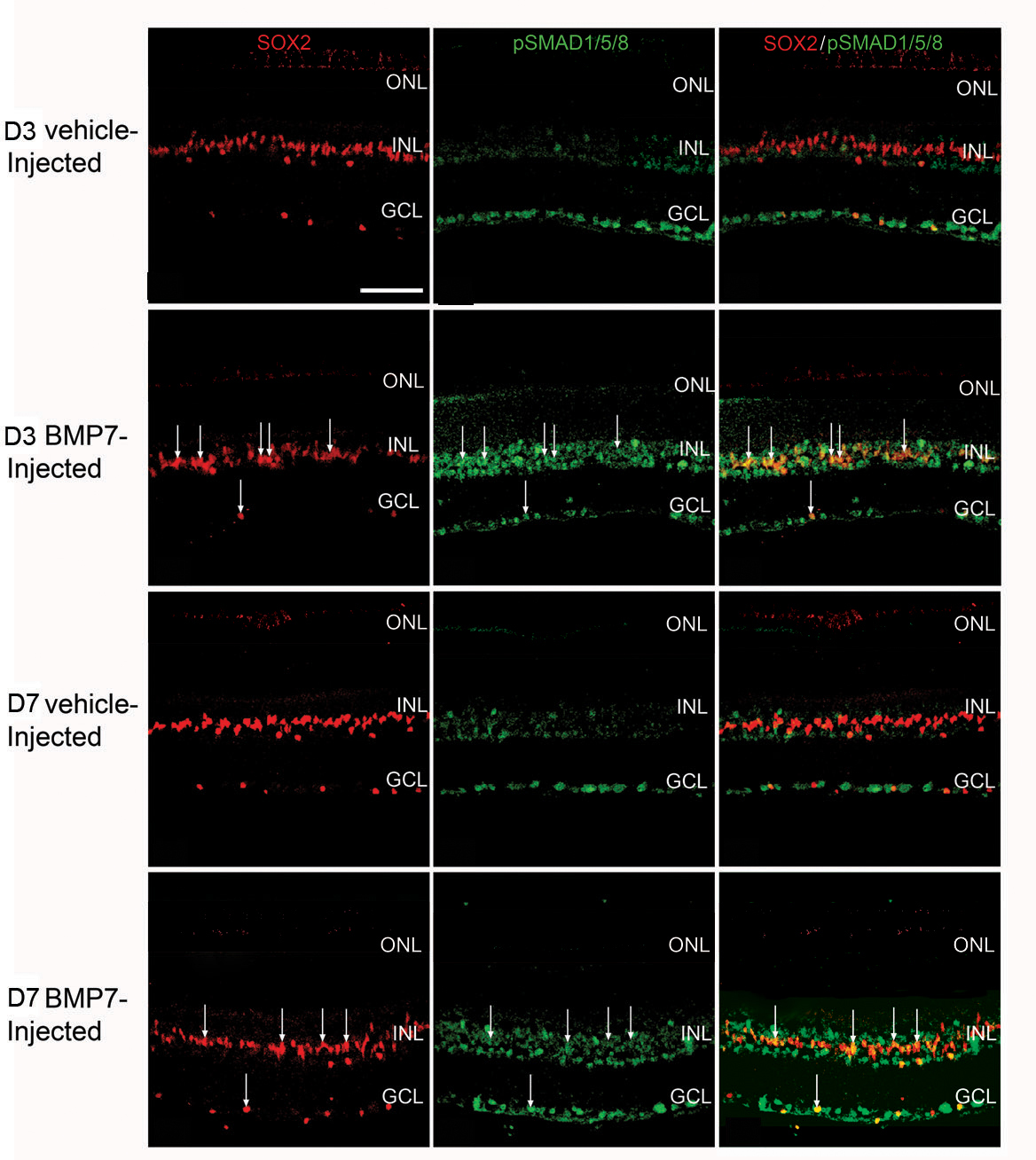
 Figure 6 of
Dharmarajan, Mol Vis 2014; 20:1085-1108.
Figure 6 of
Dharmarajan, Mol Vis 2014; 20:1085-1108.