Figure 4. Bone morphogenetic protein 7 (BMP7) treatment of MIO-M1 Müller glial cell line increases glial fibrillary acidic protein (GFAP)
expression. A: Expression patterns for a panel of markers associated with reactive gliosis were compared in human MIO-M1 Müller glial cells
treated with sodium peroxynitrite, or BMP7 for 24 or 36 h. For each experimental treatment, cells were treated with vehicle,
sodium peroxynitrite, or BMP7 and real-time quantitative PCR (RT-qPCR) was undertaken. Levels of mRNA were normalized to internal
control β2 Microglobulin, and values were plotted relative to control levels. Each sample was run in triplicate and experiment repeated 3 times for
each gene. Values represented are means±SEM. Unpaired t test was performed between the control and treated groups with * denoting a p value<0.05 and ** denoting a p value<0.005.
Any change above or below 1.0 indicates a change relative to control values. Peroxynitrite-treated MIO-M1 cells showed a statistically
significant increase above the 1.5-fold level in paired-homeobox 2 (Pax2), acid sensing ion channel 1 (Asic1), lipocalin2 (Lcn2), potassium inwardly rectifying channel 2.1 (Kir2.1), Gfap, and phosphacan (Pcan) in comparison to vehicle-treated cells. Twenty-four hours following BMP7 addition, the MIO-M1 cells only showed a statistically
significant increase above the 1.5-fold level in Gfap and Pax2, and a decrease in Txnip, galectin 3 (Gal3), Lcn2, and matrix metalloproteinase 9 (Mmp9). At the 36 h time point Gfap, and toll-like receptor 4 (Tlr4) levels of mRNA were increased above the 1.5-fold level in comparison to vehicle-treated cells and Spp1 was decreased. B: western blot analysis was performed for GFAP, S100β, TXNIP, and PAX2, with β-TUBULIN used as a loading control. Densitometric
data shown are means+/−SEM of 3 trials. Unpaired t test was performed between the control and treated groups with * denoting a p value<0.05 and ** denoting a p value<0.005.
Densitometric analysis of the blots showed a statistically significant increase in protein levels of GFAP and TXNIP in the
peroxynitrite treatment. A significant increase in levels of GFAP, S100β, and TXNIP was observed in the 24 h BMP7 treatment,
while a significant decrease was observed in PAX2 levels in the 36 h bmp7 treatment.
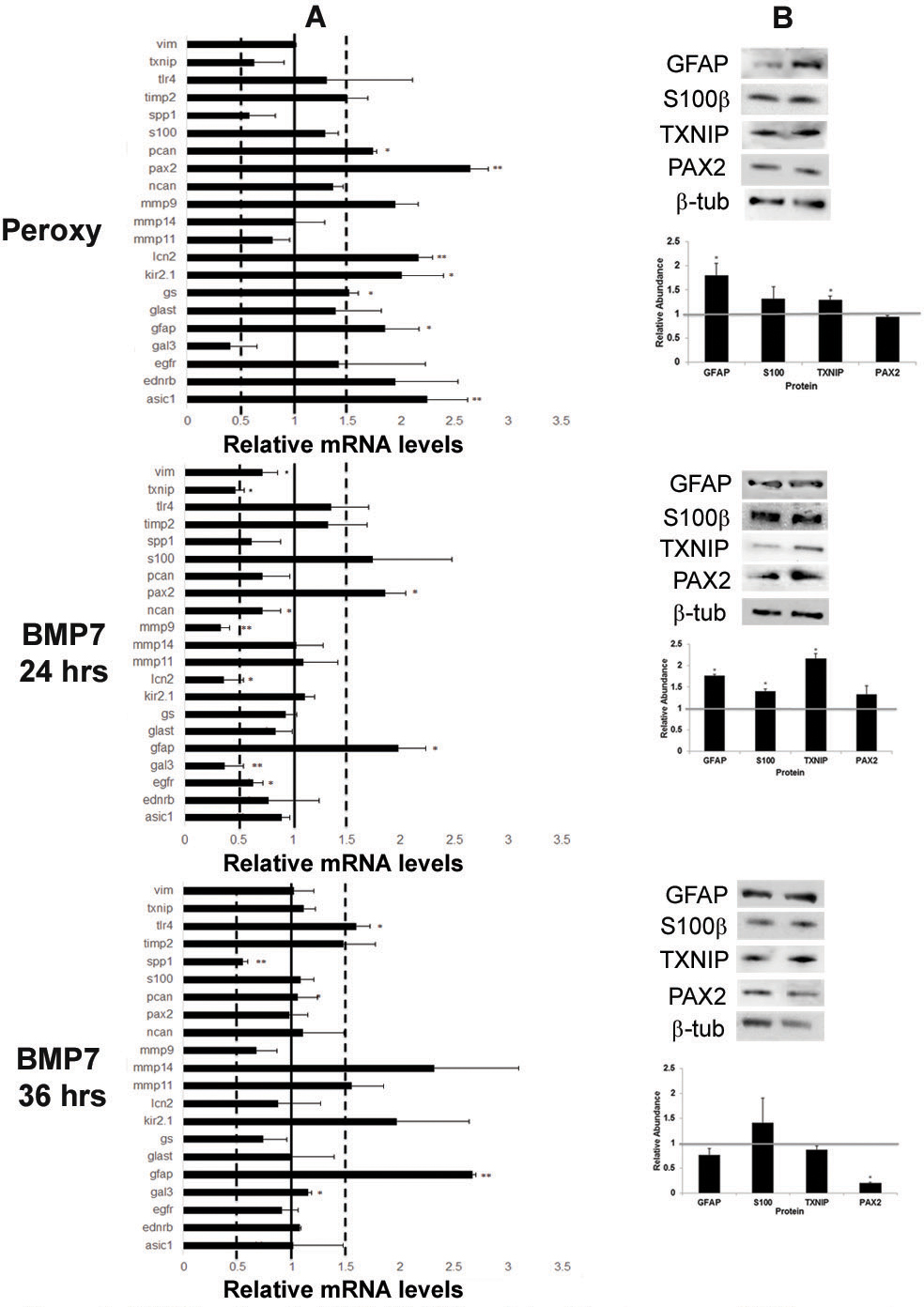
 Figure 4 of
Dharmarajan, Mol Vis 2014; 20:1085-1108.
Figure 4 of
Dharmarajan, Mol Vis 2014; 20:1085-1108.