Figure 2. Bone morphogenetic protein (BMP) signaling components in the Müller glia of adult mouse retina. A: Sections of 4 week-old retina were subjected to double-label immunofluorescence with antibodies specific for sex determining
region Y box 2 (SOX2), which labels Müller glia, retinal astrocytes, and cholinergic amacrine cells, and intracellular members
of the BMP pathway SMAD1 . SMAD1 (+) cells were localized to the inner nuclear layer (INL) and ganglion cell layer (GCL) of
P30 retina. A subpopulation of SOX2 (+) Müller glia (arrows) and retinal astrocyte cells (arrowheads) were also positive for
SMAD1. B: Sections of 4 week-old retina were subjected to double-label immunofluorescence with antibodies for SOX2, and TGF-β activated
kinase 1 (TAK1). Cells positive for the protein TAK1 were found to be localized in the INL and GCL. C: Sections of 4 week-old retina were subjected to double-label immunofluorescence with antibodies for SOX2, and FK506 rapamycin
binding protein (FKBP12). FKBP12, also part of the non-canonical BMP pathway, was found to be localized in the INL along with
sparse localization in the GCL. Very little to no co-label was seen with glutamine synthetase (GS; +) and TAK1 or FKBP12 (+)
cells (B, C). n=3 different eyes for each label. Scale Bar A=50 μm applies to all panels.
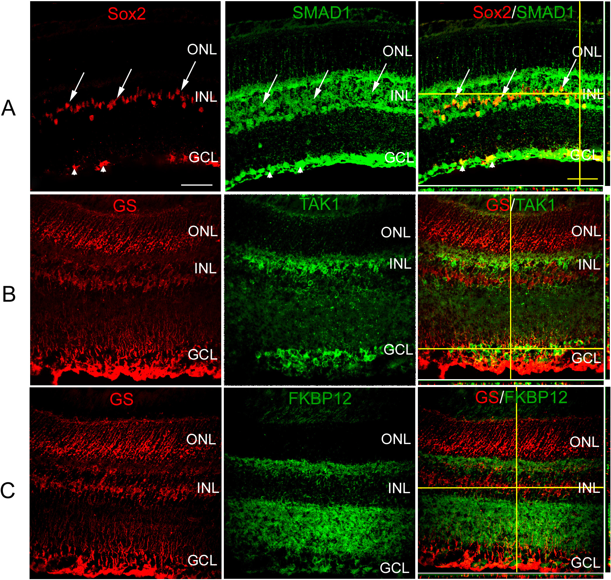
 Figure 2 of
Dharmarajan, Mol Vis 2014; 20:1085-1108.
Figure 2 of
Dharmarajan, Mol Vis 2014; 20:1085-1108.