Figure 1. Bone morphogenetic protein (BMP) type I receptors in the mature mouse retina. A: Retinal sections of 4 week-old retina double-labeled using antibodies that recognize Müller glial and retinal astrocyte
marker glutamine synthetase (GS) and BMP receptor 1A (BMPR1A). Thin plane confocal microscopy with y,z (strips to right of
panels), and x,z planes (strips at bottom of panels) are shown in the last panels in the row. BMPR1A was localized to the
Müller glial processes in the outer nuclear layer (ONL) and in Müller glial cell or retinal astrocyte processes in the ganglion
cell layer (GCL, arrows A-C). BMPR1A was also detectable in the photoreceptor outer segments, the inner plexiform layer (IPL), cell bodies within the
ganglion cell layer, and the nerve fiber layer (NFL). B: Retinal sections of 4 week-old retina double-labeled using antibodies that recognize GS and BMPR1B. BMPR1B appeared to be
localized to the outer segments (OS), outer plexiform layer (OPL) and IPL and, Müller glial cell processes as well as Müller
glial/retinal astrocyte processes in the GCL and NFL (arrows). C: Retinal sections of 4 week-old retina double-labeled using antibodies that recognize GS and activin receptor like kinase
2 (ALK2). The distribution of ALK2 receptors was similar to that of BMPR1A, with the majority of signal being localized to
the end feet within the GCL and NFL. D: Müller glia isolated from P30 retinas were co-labeled for GS and BMPR1A, 1B and ALK2. The isolated cells showed the distribution
of receptors to be primarily in the processes and the end feet of the Müller glia, as seen in the P30 tissues. n=3 different
eyes for each label. Scale bar A=50 μm applies to A, B, C.
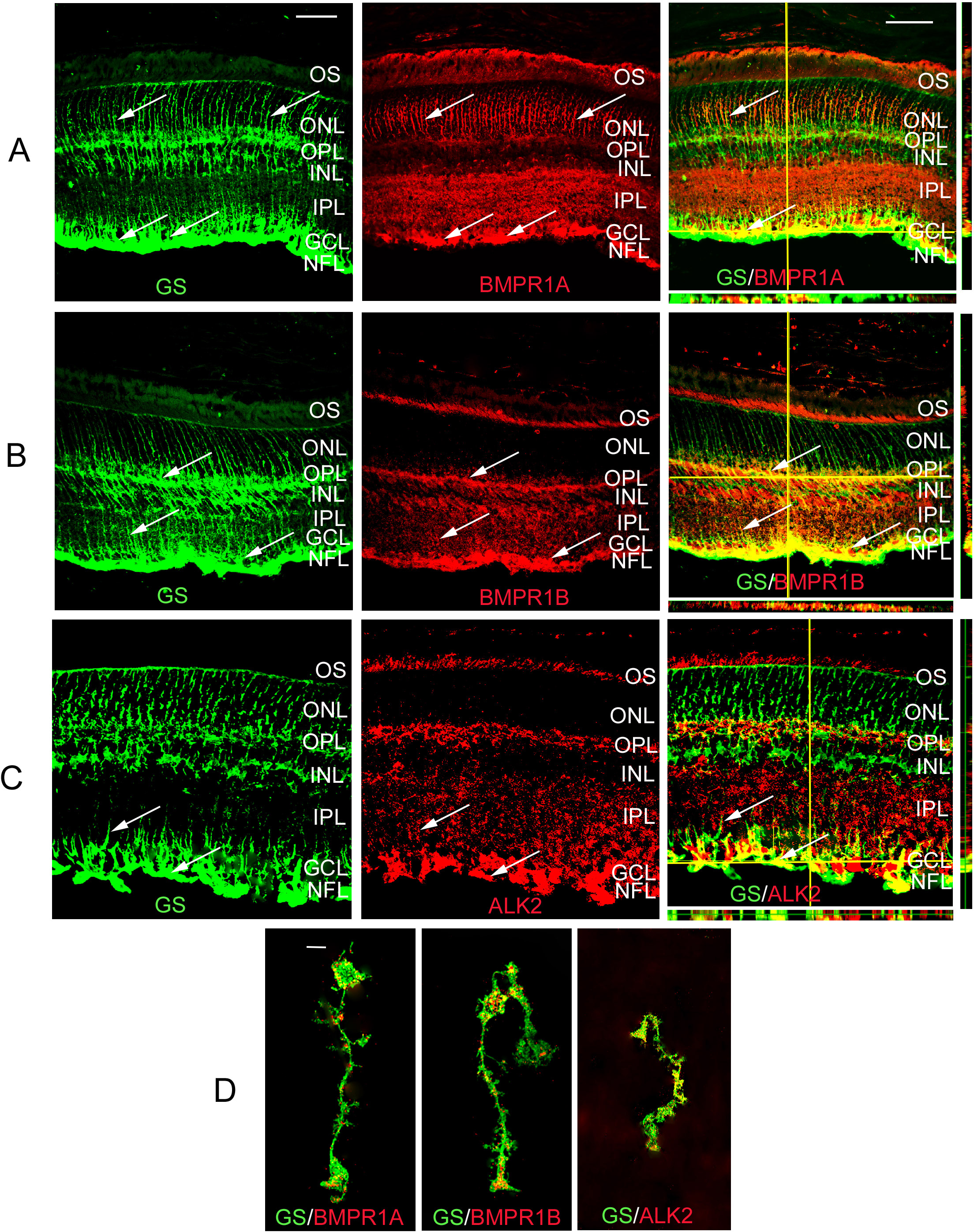
 Figure 1 of
Dharmarajan, Mol Vis 2014; 20:1085-1108.
Figure 1 of
Dharmarajan, Mol Vis 2014; 20:1085-1108.