Figure 8. Proliferating progenitor cells express tuba1a:GFP transgene following light damage. tg:1016tuba1a:GFP zebrafish were exposed to the constant intense light paradigm, and retinas were collected at the indicated time points and
assessed for green fluorescent protein (GFP, green) and proliferating cell nuclear antigen (PCNA, red) expression in the central-dorsal
retina by immunohistochemistry. Small clusters of GFP+/PCNA+ cells associate with MG processes at 51 h (double arrowheads). Large clusters of GFP+/PCNA+ cells span in the INL at 72 h (arrows). Arrowheads indicate rod precursors in the ONL. ONL, outer nuclear layer; INL, inner
nuclear layer; GCL, ganglion cell layer; IPL, inner plexiform layer. Scale bar is 50 µm.
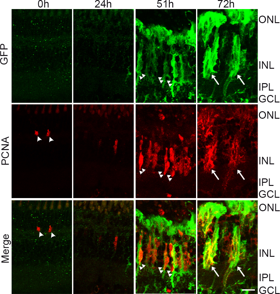
 Figure 8 of
Rajaram, Mol Vis 2014; 20:1075-1084.
Figure 8 of
Rajaram, Mol Vis 2014; 20:1075-1084.