Figure 7. Light damage triggers tuba1a:GFP transgene expression in Müller glia (MG). A: tg:1016tuba1a:GFP zebrafish were exposed to the constant intense light paradigm, and retinas were collected at the indicated time points and
assessed for GFP expression (red) by immunohistochemistry. GFP+ cells began to appear in the INL by 24 h (arrowheads). By 72 h, clusters of GFP+ cells were detected in the INL and ONL (double arrowheads). B: Immunohistochemistry for GFP and GS in 51-h light-exposed 1016tuba1a:GFP retinas. Arrows indicate GS+ and GFP+ MG. ONL, outer nuclear layer; INL, inner nuclear layer; GCL, ganglion cell layer; IPL, inner plexiform layer. Scale bars
are 50 µm.
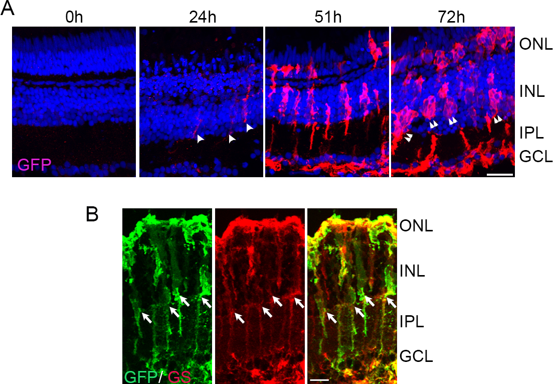
 Figure 7 of
Rajaram, Mol Vis 2014; 20:1075-1084.
Figure 7 of
Rajaram, Mol Vis 2014; 20:1075-1084.