Figure 1. Effects of constant intense light exposure on cone cells of pigmented AB zebrafish. Retinas were collected from zebrafish
before (0 h) and after the indicated length of light exposure. Retinas were processed for immunohistochemistry and stained
with zpr1 antibody (red). Nuclei were counter-stained with TOPRO (blue). Dorsal retinas are shown. The arrowheads at 0 h indicate
the ordered arrangement of the double cones; the bracket indicates ONL. White arrows show condensed zpr+ cells at 72 h of intense light. ONL, outer nuclear layer; INL, inner nuclear layer; GCL, ganglion cell layer. Scale bar is
50 µm.
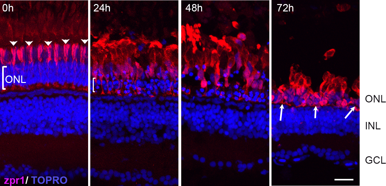
 Figure 1 of
Rajaram, Mol Vis 2014; 20:1075-1084.
Figure 1 of
Rajaram, Mol Vis 2014; 20:1075-1084.