Figure 1. Representative immunostaining results for β-catenin. A: The negative control has the first antibody replaced with immunoglobulin. B: β-catenin protein expression detected in the membrane (200×), and (C) aberrant localization of β-catenin in the cytoplasm/nuclei (200×).
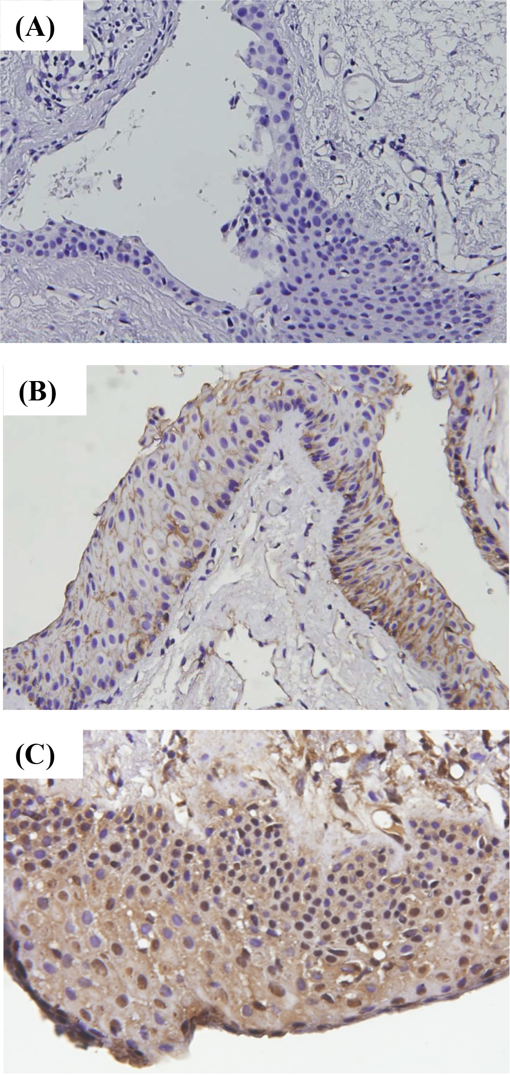
 Figure 1 of
Wu, Mol Vis 2014; 20:1048-1056.
Figure 1 of
Wu, Mol Vis 2014; 20:1048-1056.