Figure 6. Far ultraviolet circular dichroism spectroscopy. The far ultraviolet circular dichroism (UV–CD) spectra of gamma irradiation
and non-gamma irradiation αA-crystallin wild-type and N-terminal mutants, R12C, R21L, R49C, and R54C, were determined with
CD spectroscopy. The secondary structure profile of the αA-crystallin wild-type (wt) and N-terminal mutants was evaluated
at (A) 0 kGy, (B) 0.5 kGy, (C) 1.0 kGy, and (D) 2.0 kGy dosages of GI. The secondary structure profile of the gamma irradiated (GI) αA-crystallin wt was stable at different
dosages. However, significant changes in the β-sheet and random coil content in the GI mutants, R12C, R21L, and R49C, was
observed at 0.5 kGy. The mutant R54C exhibited changes in β-sheet content only after 1.0 kGy of GI. Each spectrum is an average
of five runs. The spectra were recorded between 195 and 250 nm.
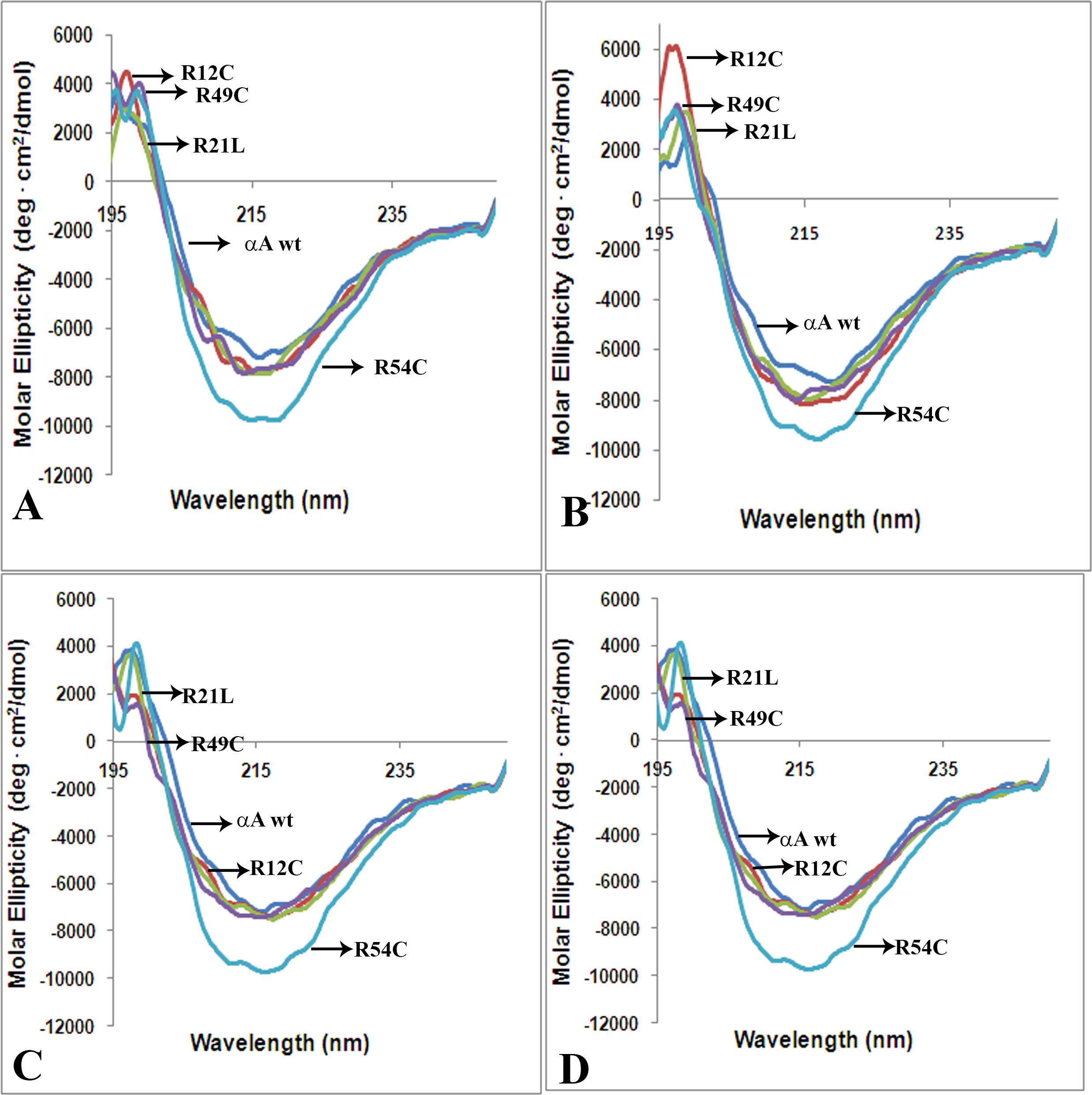
 Figure 6 of
Ramkumar, Mol Vis 2014; 20:1002-1016.
Figure 6 of
Ramkumar, Mol Vis 2014; 20:1002-1016.