Return to Non-Uniform Distribution of the NMDAR1...
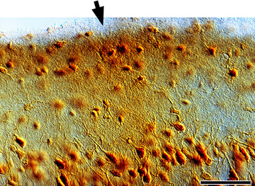
Figure 6. NMDAR1 immunoreactivity in layer 1 and the superficial aspect of
layer 2 in kitten visual cortex.
Within an NMDAR1 patch (filled arrow) there is
a dense mesh of fine NMDAR1 positive processes in layer 1 and darker neuropil
label. Scale bar is 50”m.