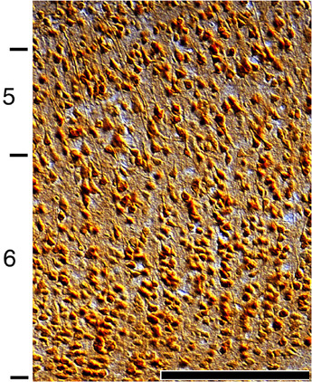
Return to Non-Uniform Distribution of the NMDAR1...
Figure 5. Light micrograph of a coronal section through layers 5 and 6 of
kitten area 17.
NMDAR1 labelled cell bodies are present in both layers 5 and 6
with many NMDAR1 positive cells in the deeper half of layer 6. Scale bar is
100”m.