Return to Non-Uniform Distribution of the NMDAR1...
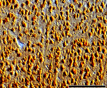
Figure 4. NMDAR1 label in layer 4 of kitten visual cortex at 4 weeks of age.
At
this age there are many NMDAR1 positive neurons in the geniculocortical
recipient layer and the staining is darker in the ventral half of the layer.
Some labeled segments of processes, probably apical dendrites from
infragranular pyramidal neurons, are present. Scale bar 50”m.