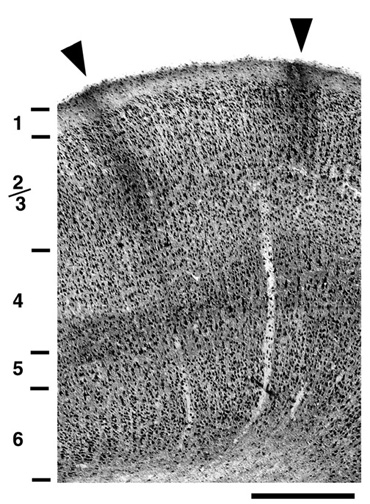
 This light micrograph from a coronal section shows the full thickness of the visual cortex
and the laminar variation in labelling. Layers were determined from an adjacent
Nissl stained section. In the superficial layers there are two prominent dark
patches of NMDAR1 immunoreactivity (filled arrows) that extend from layer 1
down to the top of layer 4. Scale bar is 500”m.
This light micrograph from a coronal section shows the full thickness of the visual cortex
and the laminar variation in labelling. Layers were determined from an adjacent
Nissl stained section. In the superficial layers there are two prominent dark
patches of NMDAR1 immunoreactivity (filled arrows) that extend from layer 1
down to the top of layer 4. Scale bar is 500”m.