![]() Figure 5 of Yeagle et al.
Figure 5 of Yeagle et al.
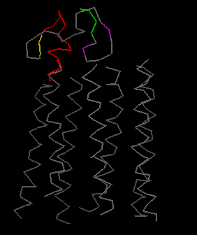
Figure 5. Animated docking of the average structure for the carboxyl terminal (rhoIVe) of rhodopsin
Average of the six best structures (shown in Figure 4) for the carboxyl terminal (rhoIVe) of rhodopsin. In the animation, the carboxyl terminal (rhoIVe) of rhodopsin is initially shown above (and separated from) helix 7 of the transmembrane domain of rhodopsin. The molecule is translated downward to show how it docks to helix 7 and then the two moieties are rotated together about two axes to display the spatial relationships of this structure. Regions of the carboxyl terminal (rhoIVe) of rhodopsin are also color coded to demonstrate various features of the structure.
Note that the slide bar at the bottom of the quicktime movie can be used to manually control the flow of the movie. If you do not want to or are unable to view the movie, a representative frame is included below as a still image.
| Color | Feature |
|---|---|
| Purple | Two most readily phosphorylated serines |
| Yellow | Two cysteines that can be palmitoylated form one end of the fourth cytoplasmic loop |
| Red | Fourth cytoplasmic loop |
| Green | Mutation sites on the carboxyl terminal of rhodopsin associated with retinitis pigmentosa |