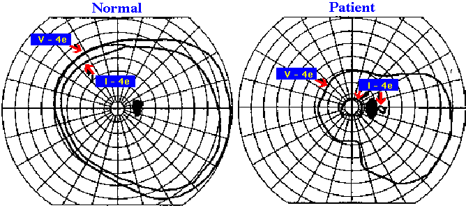
The kinetic fields are for the right eye in each subject using both V-4e and I-4e test targets.
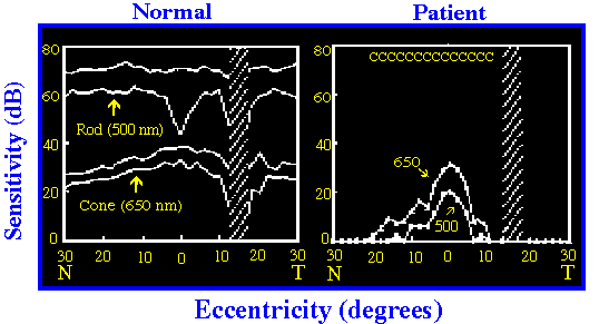
For the normal, the lines delimit +/- 2 standard deviations from the mean normal using the 500 nm stimulus (rod-mediated detection, upper set) and a 650 nm stimulus at the cone plateau (cone-mediated detection, lower set). For the patient profiles, the curves describe the responses to the two different stimulus wavelengths used. Letters above the sensitivity measurements in the patient are the photoreceptor mediations for detection of the 500 and 650 nm stimuli: C = cone detection. N = nasal; T = temporal.
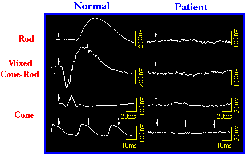
Arrows indicate stimulus onset and calibrations are to the right and
below the responses.