Figure 10. Confocal microscopy of sCD44-treated human trabecular meshwork cells. A: sCD44-treated cells were stained with rhodamine phalloidin (red) and FITC anti-annexin 2 (green). B: sCD44-treated cells were stained with rhodamine phalloidin and FITC anti-phosphorylated annexin 2. Noticeable structural
differences were observed in the samples treated with 0.1 ng sCD44 compared to PBS controls. There was also an observed increase
in the concentration of phosphorylated annexin 2 surrounding the spokes and vertices of the cross-linked actin network formations.
This co-localization of F-actin and phosphorylated annexin 2 can be seen when images of the specific fluorescence of each
substance were overlapped spatially on their precise axes. Magnification is 200×.
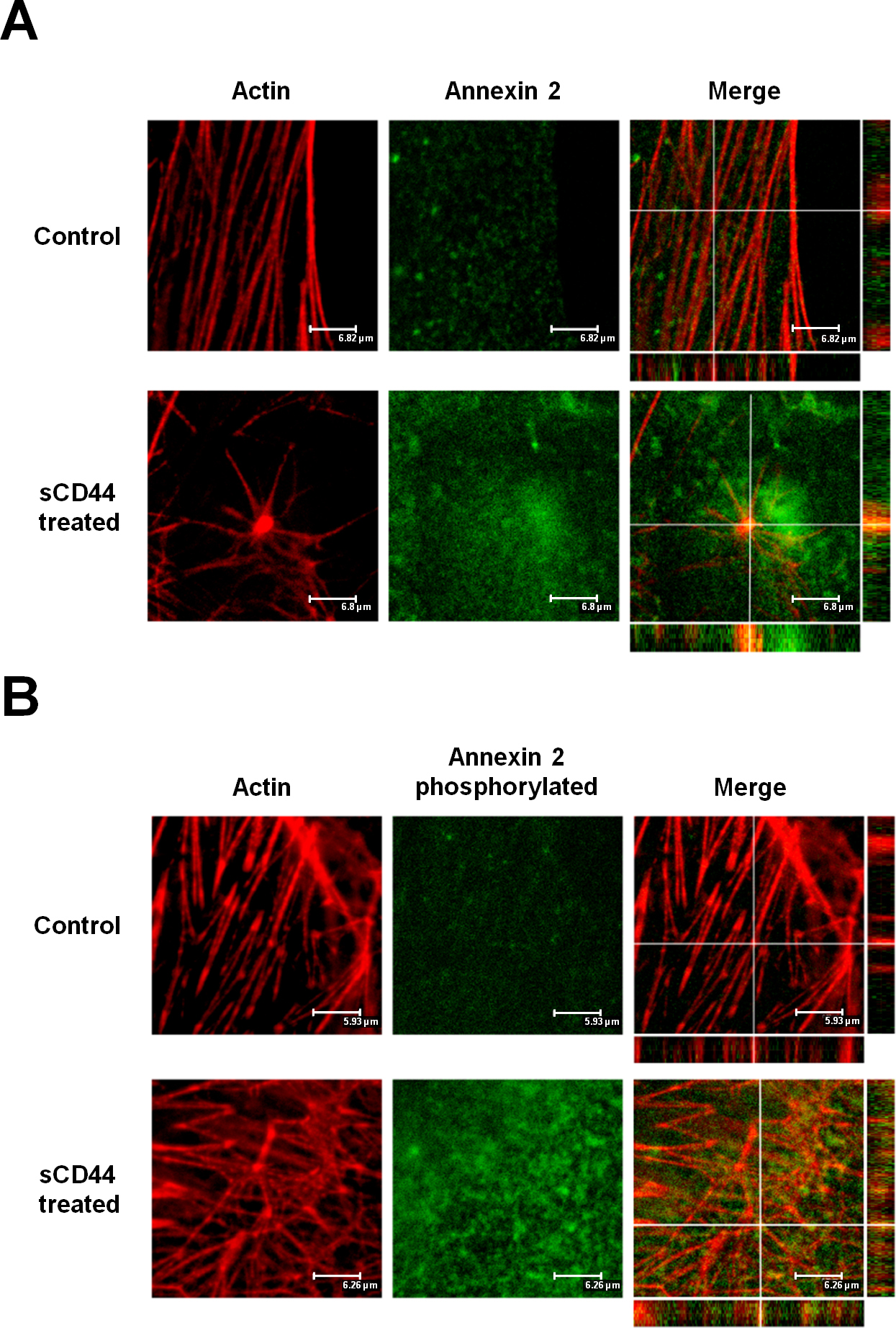
 Figure 10 of
Giovingo, Mol Vis 2013; 19:2151-2164.
Figure 10 of
Giovingo, Mol Vis 2013; 19:2151-2164.