Figure 7. The deposition of type III collagen and accumulation of myofibroblasts in the cornea after alkali injury. The expression of
type I (A, D, G) and III (B, E, H) collagen and α-smooth muscle actin (α-SMA, C, F, I) in the corneas in normal (A–C), vehicle-treated (D–F), and peroxisome proliferator-activated receptor gamma-treated (G–I) corneas (A, D, G: type I collagen stain; B, E, H: type III collagen stain; C, F, I: α-SMA stain, scale bar: 100 μm) on day 14. Type I collagen was the main collagen expressed in the cornea, and the deposition
of type III collagen and accumulation of α-SMA-positive myofibroblasts was noted in the injured cornea, but was deposited
to a lesser degree in the peroxisome proliferator-activated receptor gamma (PPARγ) group than in the vehicle group.
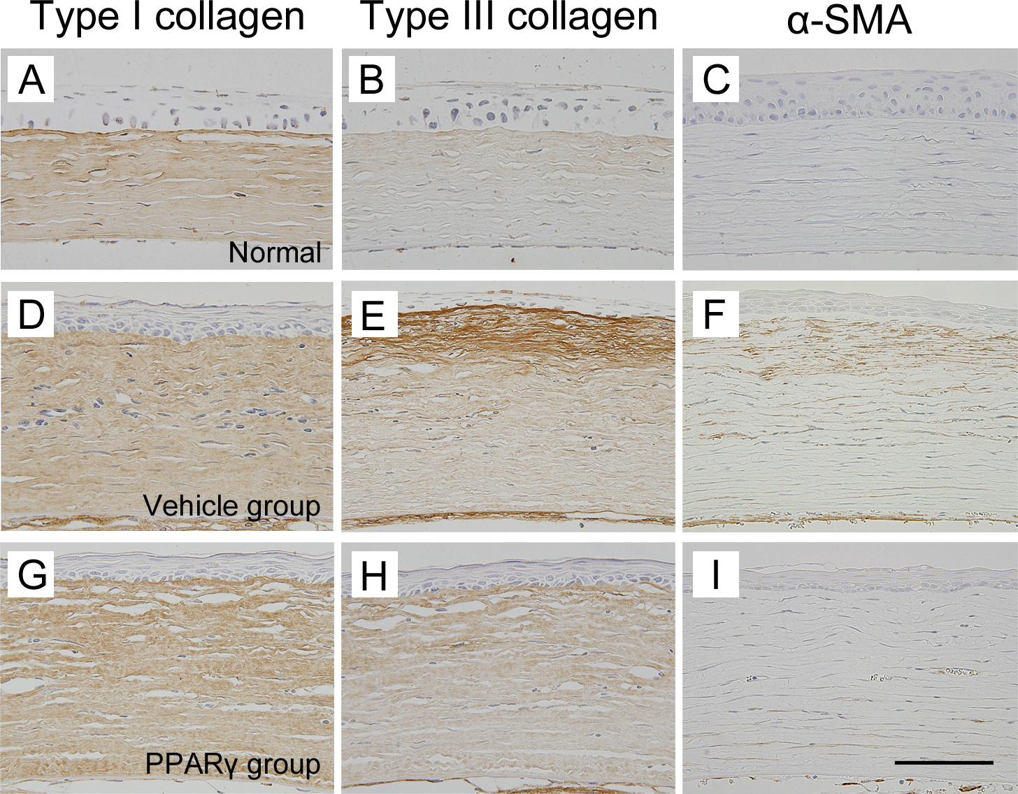
 Figure 7 of
Uchiyama, Mol Vis 2013; 19:2135-2150.
Figure 7 of
Uchiyama, Mol Vis 2013; 19:2135-2150.