Figure 5. The infiltration of M2 macrophages in the alkali-burned cornea. Representative photomicrographs of infiltrating M2 macrophages
in the vehicle (A, C) and peroxisome proliferator-activated receptor gamma (PPARγ) (B, D) groups on day 1 (A, B) and day 7 (C, D) after alkali injury (A–D: ED2 stain, scale bar: 100 μm) showed that, in the vehicle and PPARγ groups, the infiltration of ED2-positive M2 macrophages
was prominent on day 1 in the peripheral regions of the cornea and on day 7 in the central regions of the cornea. The degree
of M2 macrophage infiltration was more prominent in the PPARγ group than in the vehicle group. E: The number of ED2-positive M2 macrophages per 400X high-power fields in the cornea showed that the infiltration of M2 macrophages
in the cornea was significantly increased in the PPARγ group compared to the vehicle group. F: The percentage of ED2-positive M2 macrophages in the total ED1-positive macrophages in the cornea showed that the percentage
of M2 macrophages in the total macrophage population was increased more in the PPARγ group compared to the vehicle group.
The results are presented as the means±standard errors. *p<0.05, **p<0.01, compared with the vehicle group. In the double
immunofluorescence studies with ED2 (red; M2 marker; G, J) and ED3 (green; activated macrophage marker; H, K) in the vehicle and PPARγ groups on day 7, the ED2-positive M2 macrophages were more prominent than the ED3-positive activated
macrophages in the PPARγ group (J–L, 800X), although the ED3-positive cells were more prominent than the ED2-positive cells in the vehicle group (G–I, 800X).
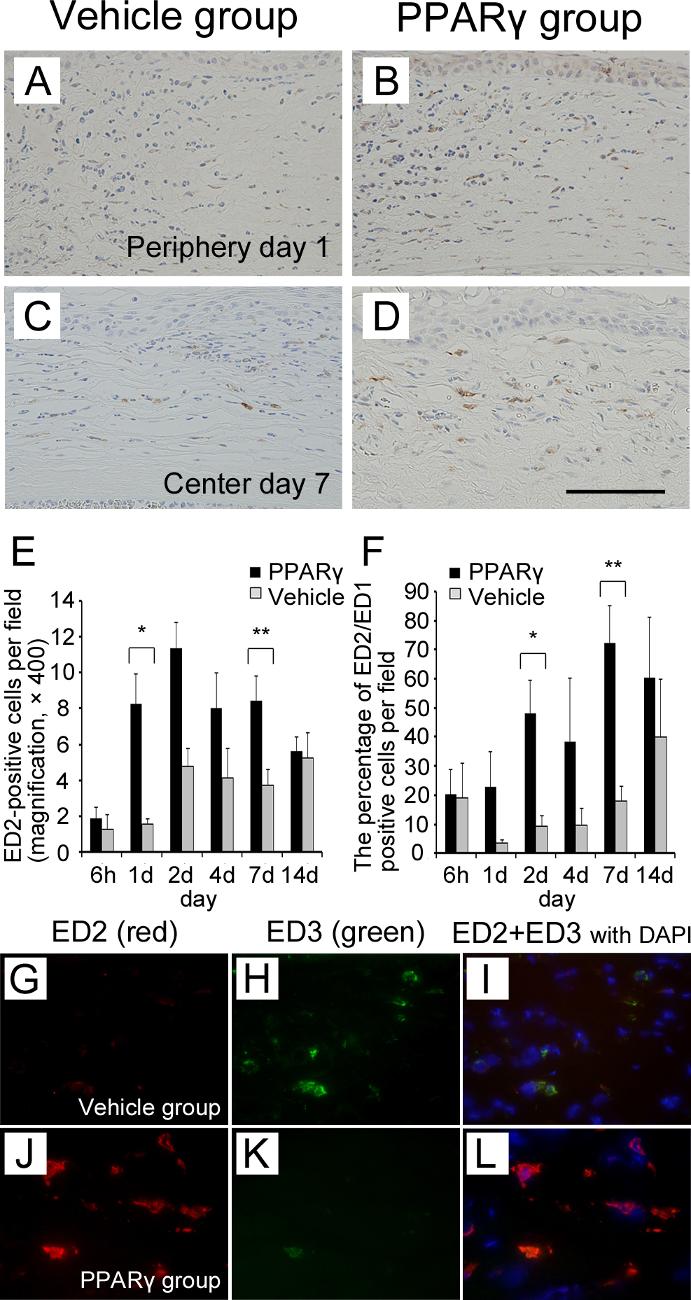
 Figure 5 of
Uchiyama, Mol Vis 2013; 19:2135-2150.
Figure 5 of
Uchiyama, Mol Vis 2013; 19:2135-2150.