Figure 4. The infiltration of ED1-positive macrophages in alkali-burned corneas. Representative photomicrographs of infiltrating macrophages
in the vehicle (A, C) and peroxisome proliferator-activated receptor gamma (PPARγ) (B, D) groups on day 1 (A, B) and day 7 (C, D) after alkali injury (A–D: ED1 stain, scale bar: 100 μm) showed that, in the vehicle and PPARγ groups, the macrophage infiltration was prominent on
day 1 in the peripheral regions of the cornea and on day 7 in the central regions of the cornea. The degree of macrophage
infiltration was less severe in the PPARγ group than in the vehicle group on day 1 and day 7. E: The number of ED1-positive macrophages per 400X high-power fields in the cornea showed that the macrophage infiltration
in the cornea was significantly inhibited in the PPARγ group compared to the vehicle group on days 1, 2, and 7 in the alkali-burned
cornea. The results are presented as the means±standard errors. *p<0.05, **p<0.01, compared with the vehicle group.
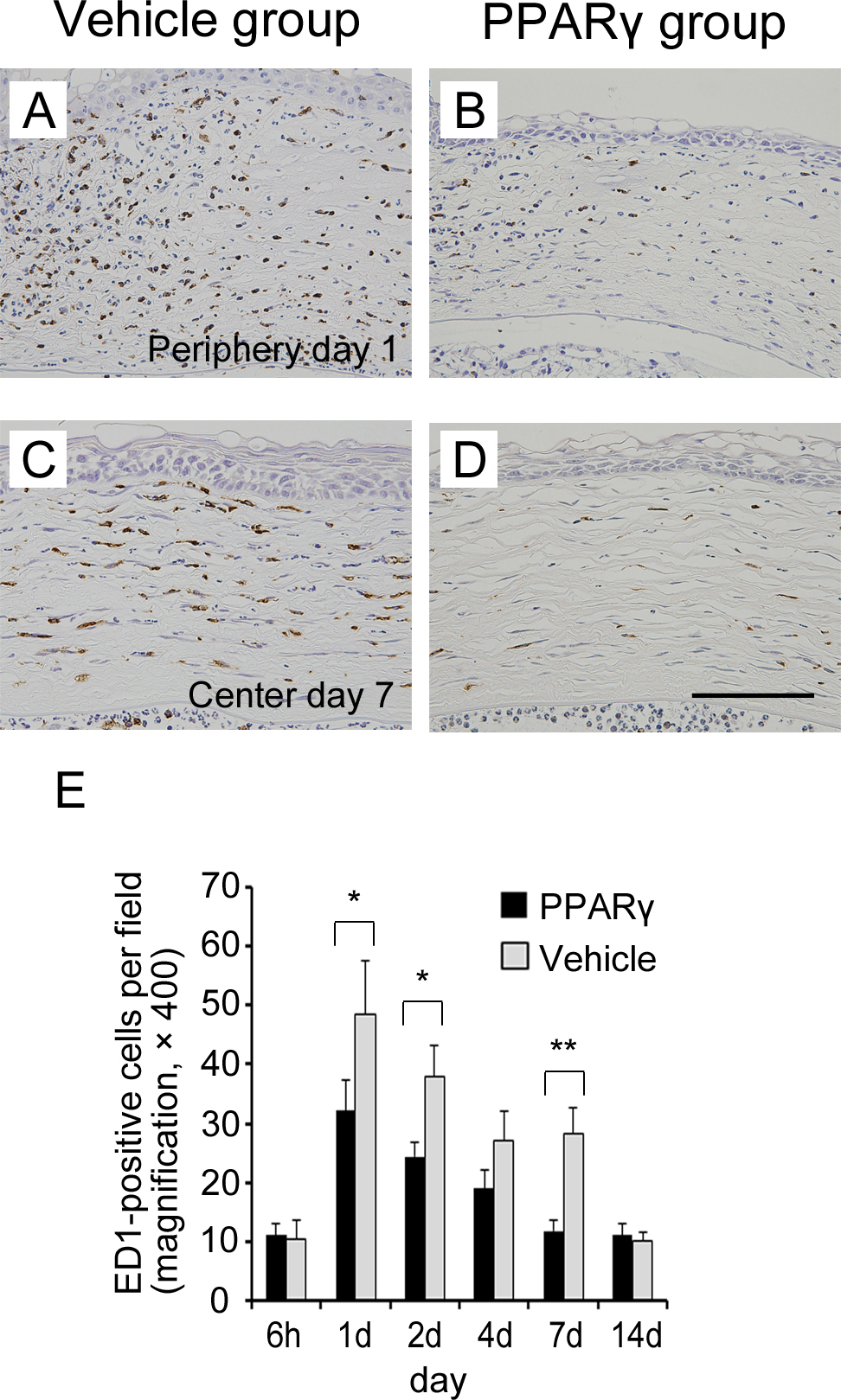
 Figure 4 of
Uchiyama, Mol Vis 2013; 19:2135-2150.
Figure 4 of
Uchiyama, Mol Vis 2013; 19:2135-2150.