Figure 2. The corneal wound healing after alkali burn injury. The development of corneal wound healing after alkali injury in the vehicle
(A–D: peripheral regions, I–L: central regions) and peroxisome proliferator-activated receptor γ (E–H: peripheral regions, M–P: central regions, scale bar: 100 μm) groups. After alkali injury in the vehicle and peroxisome proliferator-activated receptor
gamma (PPARγ) groups, various inflammatory cells infiltrated from the corneal limbus into the corneal center by day 7. Between
6 h and day 1 after the injury, the inflammatory cells were prominent in the peripheral regions of the cornea, and were increased
in the central regions of the cornea on day 7. After inflammatory cell infiltration, corneal neovascularization developed
from the corneal limbus to the center by day 7 to day 14. The severity of the inflammatory cell infiltration and the degree
of neovascularization in the cornea were decreased in the PPARγ group compared to the vehicle group.
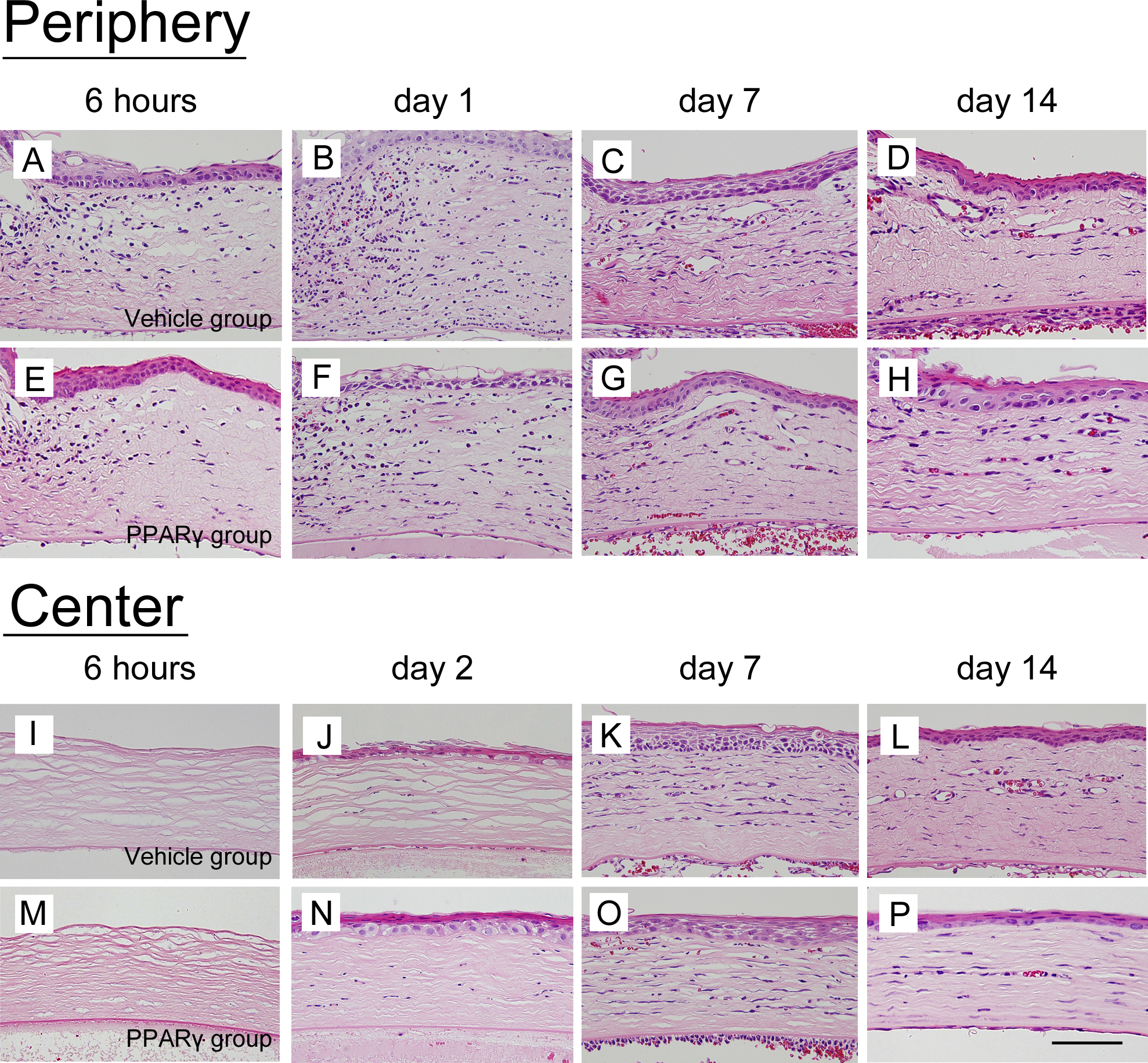
 Figure 2 of
Uchiyama, Mol Vis 2013; 19:2135-2150.
Figure 2 of
Uchiyama, Mol Vis 2013; 19:2135-2150.