Figure 1. Comparison of the retina of 4Bnr-BALB/c-Rd3rd3/rd3 (rd3−/−) and 4Bnr-BALB/c-Rd3+/+ (rd3+/+) at P21. A: The nuclear layers stained with 4', 6-diamidino-2-phenylindole (blue) revealed that the outer nuclear layer (ONL) of rd3−/− is thinner than that of wild-type. Labeling with the antibody to GC1 (green) showed that GC1 is localized to the outer segments
of the wild-type retina but is not detectable in the rd3−/− retina. In the control experiment where only the anti-mouse secondary antibody (Alexa 488) was used, punctate labeling in
the OPL and the IPL was observed. The secondary antibody was likely labeling the retinal vasculature. B: The ONL thickness of rd3−/− was reduced by 42±4% compared to wild-type. Error bars represent the standard deviation of the mean (n=4). Abbreviations:
RPE, retinal pigmented epithelium; OS, outer segment; IS, inner segment; ONL, outer nuclear layer; OPL, outer plexiform layer;
INL, inner nuclear layer; IPL, inner plexiform layer; GCL, ganglion cell layer.
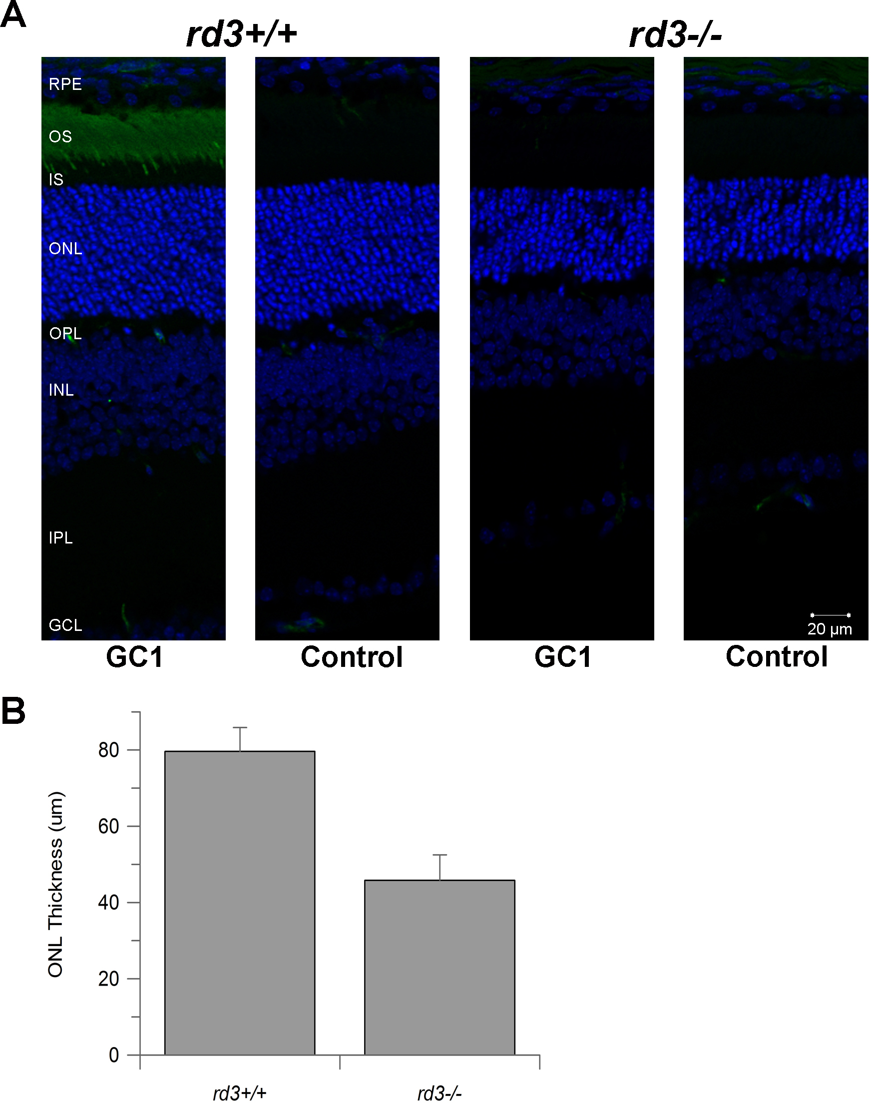
 Figure 1 of
Cheng, Mol Vis 2013; 19:955-969.
Figure 1 of
Cheng, Mol Vis 2013; 19:955-969.