Figure 5. Ocular characteristics and systemic anomalies of patient II:3. Slit lamp photographs showed hypoplasia and iris posterior
embryotoxon (black arrows) in both eyes, but Haab’s striae were observed only in the left eye. A, B: Gonioscopy showed open angles in both eyes with anterior insertion of the iris into the trabecular meshwork, prominent iris
processes, and broad-based synechiae at places in both eyes (C, D). Physical examination revealed hypertelorism, telecanthus, a flat face, and a flat broad nasal bridge (E, F).
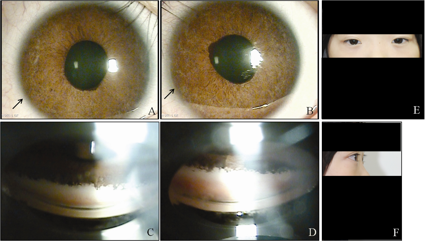
 Figure 5 of
Kim, Mol Vis 2013; 19:935-943.
Figure 5 of
Kim, Mol Vis 2013; 19:935-943.