Figure 4. Ocular characteristics and systemic anomalies of patient II:2. Slit lamp photographs showed iris hypoplasia and posterior
embryotoxon (black arrows) in both eyes, but Haab’s striae were observed only in the right eye (A, B). Gonioscopy showed open angles in both eyes with anterior insertion of the iris into the trabecular meshwork, prominent
iris processes, and broad-based synechiae at places in both eyes (C, D). Physical examination revealed hypertelorism, telecanthus, a flat face, and a flat broad nasal bridge (E, F).
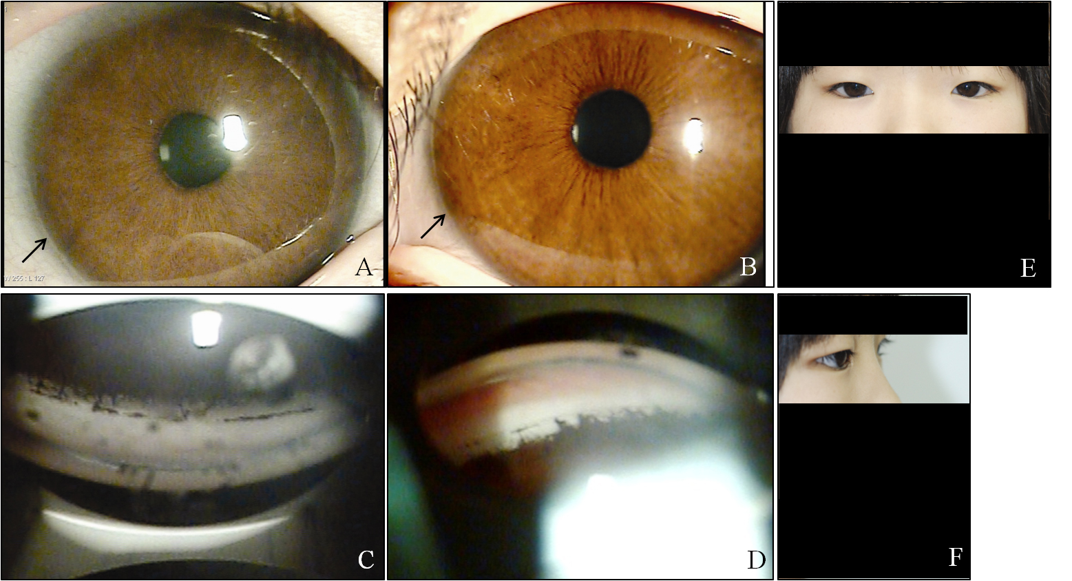
 Figure 4 of
Kim, Mol Vis 2013; 19:935-943.
Figure 4 of
Kim, Mol Vis 2013; 19:935-943.