Figure 3. Ocular characteristics and systemic anomalies of patient I:1. Slit lamp photographs showed slight corneal edema, Haab’s striae,
iridocorneal adhesions, and atrophy of the iris with corectopia in both eyes (A, B). Gonioscopy showed open angles in both eyes with anterior insertion of the iris into trabecular meshwork, prominent iris
processes, and broad-based synechiae at places in both eyes (C, D). Physical examination revealed exotropia in the left eye, a flat midface, hypertelorism, and telecanthus (E, F).
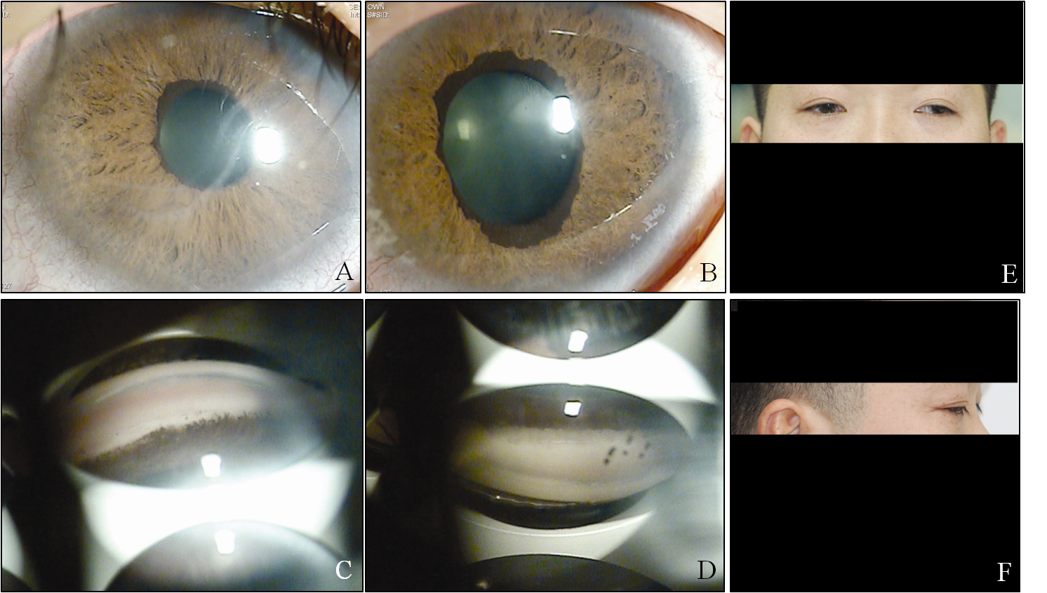
 Figure 3 of
Kim, Mol Vis 2013; 19:935-943.
Figure 3 of
Kim, Mol Vis 2013; 19:935-943.