Figure 1. Comparison of the effects of hydrogen peroxide at various concentrations on the viability of orbital fibroblasts between patients
with Graves’ ophthalmopathy (GO) and normal controls. We treated orbital fibroblasts from patients with GO (n=7) and age-matched
normal subjects (n=5) with various concentrations of hydrogen peroxide (H2O2) for 24 h. The cell proliferation rate was examined with the AlamarBlue assay as methods described and normalized to each
control not exposed to H2O2. The mean values of cell proliferation in H2O2-treated orbital fibroblasts are shown in the histogram. Treatment with a low concentration of H2O2 (<25 μM) in GO orbital fibroblasts significantly induced the cell proliferation, but the effect was not observed in normal
subjects. The cytotoxicity of H2O2 was observed in GO orbital fibroblasts above 50 μM and in normal subjects above 100 μM. The data are presented as mean ±
standard deviation of the results from three independent experiments. (Significant increase when ** p<0.01 and *p<0.05; significant
decrease when ##p<0.01 and #p<0.05.)
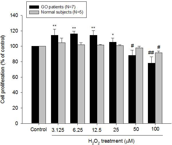
 Figure 1 of
Tsai, Mol Vis 2013; 19:927-934.
Figure 1 of
Tsai, Mol Vis 2013; 19:927-934.