Figure 4. Isolated bipolar cells from wild-type and C-terminal-binding protein 1(CtBP1)-knockout retinas have similar numbers of ribbons
marked with RIBEYE-binding peptide. A: The panels show confocal images of a live synaptic terminal loaded with RIBEYE-binding peptide in wild-type (WT; left) and
CtBP1-knockout (KO; right) bipolar neurons. The peptide was dialyzed into cells via a whole-cell patch pipette. B: The average number of ribbons per terminal was not significantly different (p=0.22) between WT (n=10) and CtBP1-KO (n=11)
bipolar neurons. The error bars indicate ±one standard error of the mean.
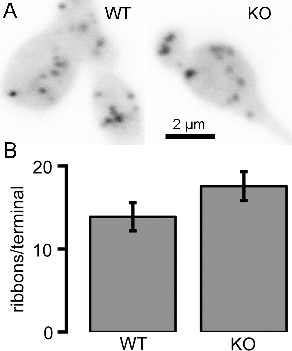
 Figure 4 of
Vaithianathan, Mol Vis 2013; 19:917-926.
Figure 4 of
Vaithianathan, Mol Vis 2013; 19:917-926.