Figure 2. Overall morphology of the retina and localization of synaptic ribbons are normal in C-terminal-binding protein 1 (CtBP1)-knockout
(KO) mice. A: The panel shows examples of confocal images obtained from transverse sections of wild-type (WT) and CtBP1-KO retina stained
with an antibody against C-terminal-binding protein 2 (CtBP2) to reveal ribbons. The intensity scale is inverted for better
visualization. ONL refers to the outer nuclear layer, OPL indicates the outer plexiform layer, INL denotes the inner nuclear
layer, IPL points to the inner plexiform layer, and GCL specifies the ganglion cell layer. B: The panel shows a higher magnification confocal image of the OPL, containing the crescent-shaped ribbons of rod photoreceptors.
C: The panel shows a higher magnification confocal image of sublamina b of the inner plexiform layer (IPLb), demonstrating the typical punctate ribbons of bipolar neurons.
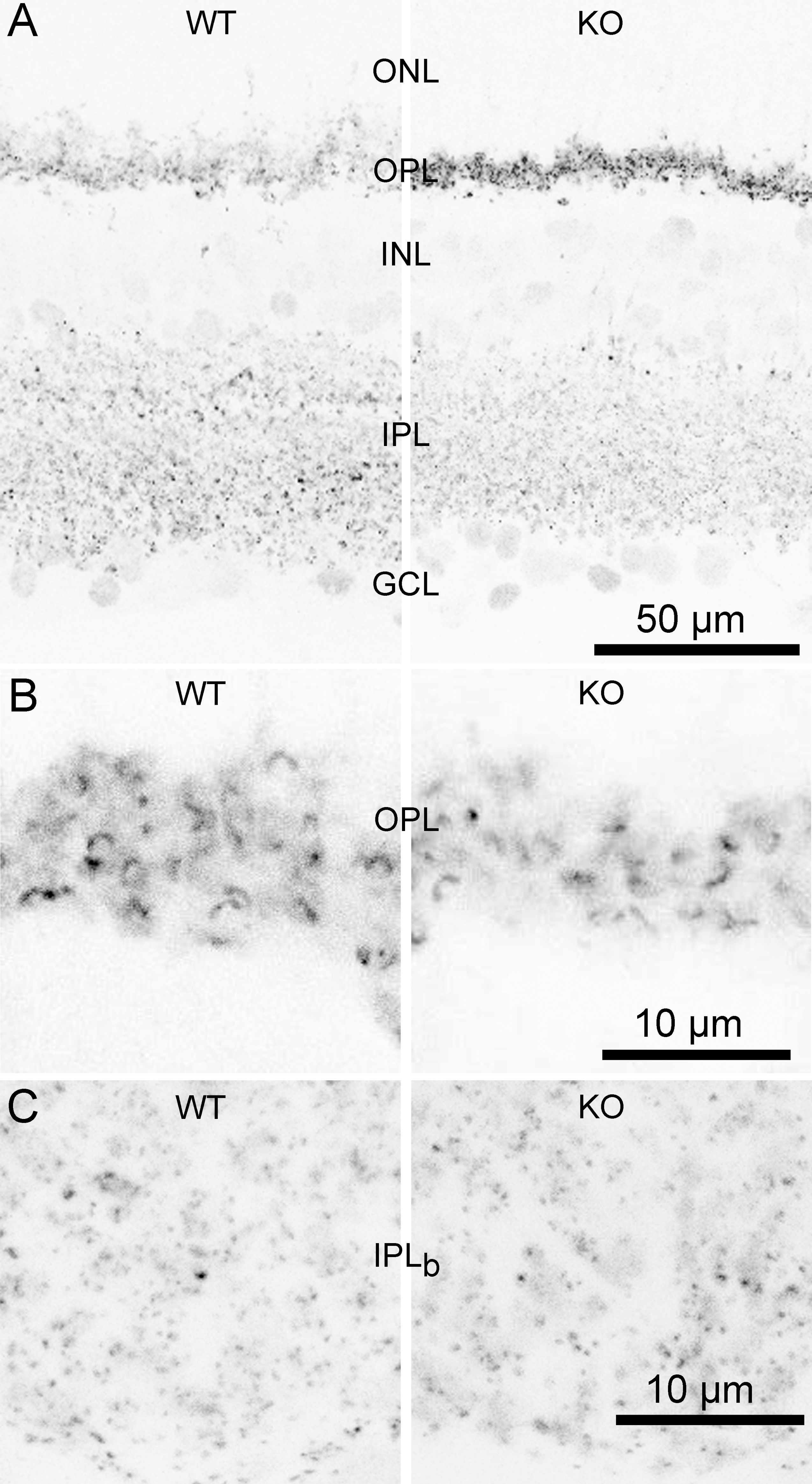
 Figure 2 of
Vaithianathan, Mol Vis 2013; 19:917-926.
Figure 2 of
Vaithianathan, Mol Vis 2013; 19:917-926.