Figure 1. C-terminal-binding protein 1 (CtBP1) immunostaining at ribbon synapses is absent in CtBP1-knockout mouse retina. A: The panel shows an example of a confocal image obtained from a transverse section of WT mouse retina stained with a mouse
monoclonal antibody against CtBP1. The intensity scale is inverted, so that brighter spots appear in black. ONL refers to
the outer nuclear layer, OPL indicates the outer plexiform layer, INL denotes the inner nuclear layer, IPL points to the inner
plexiform layer, and GCL specifies the ganglion cell layer. B: The panel shows an example of a confocal image obtained from a transverse section of CtBP1-knock out (KO) retina stained
with anti-CtBP1. Intensity scale is inverted. Staining in the plexiform and nuclear layers was similar to that in sections
stained with the anti-mouse secondary antibody alone, without anti-CtBP1, indicating the absence of CtBP1 in KO retina. The
scattered dark spots (bright staining) were also present in sections stained with the secondary antibody alone and likely
reflect blood vessels. Scale bar in B applies to A as well.
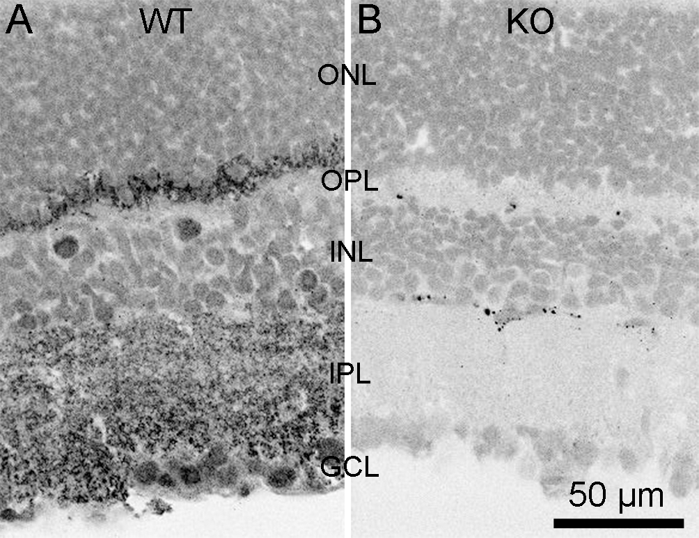
 Figure 1 of
Vaithianathan, Mol Vis 2013; 19:917-926.
Figure 1 of
Vaithianathan, Mol Vis 2013; 19:917-926.