Figure 2. Retinal cells labeled with the lectin IB4 from Griffonia simplicifolia (IB4, green) in fresh neuroretinal samples (A) and experimental 9-day cultures (B–E). Ada: adalimumab treated culture; DAPI: 4’,6-diamino-2-phenylindole dihydrochloride staining (blue); INL: inner nuclear
layer; GCL: ganglion cell layer; ONL: outer nuclear layer; TNFα: tumor necrosis factor alpha treated culture; TNFα/Ada: tumor
necrosis factor alpha plus adalimumab treated culture. 4’,6-diamino-2-phenylindole dihydrochloride (DAPI) staining (blue)
was present in the nuclei of the ganglion cell layer (GCL), the inner nuclear layer (INL), and the outer nuclear layer (ONL).
IB4-labeled cells were present between the INL and the GCL and extended into the INL in the fresh samples and the 9-day culture
experiments (A–E, arrows). Scale bars: 20 µm.
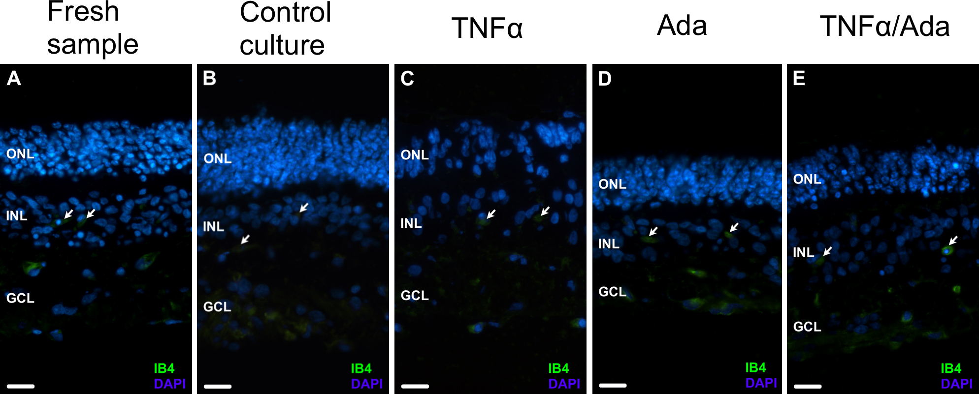
 Figure 2 of
Fernandez-Bueno, Mol Vis 2013; 19:894-903.
Figure 2 of
Fernandez-Bueno, Mol Vis 2013; 19:894-903.