Figure 1. Retinal distribution of glial fibrillary acidic protein (GFAP, red; A, D, G, J, M) and cellular retinaldehyde-binding protein (CRALBP, green; B, E, H, K, N), and corresponding merged compositions (Merge; C, F, I, L, O), in fresh neuroretinal samples (A–C) and experimental 9 day culture conditions (D–O). Ada: adalimumab treated culture; DAPI: 4’,6-diamino-2-phenylindole dihydrochloride staining (blue); INL: inner nuclear
layer; GCL: ganglion cell layer; ONL: outer nuclear layer; TNFα: tumor necrosis factor alpha treated culture; TNFα/Ada: tumor
necrosis factor alpha plus adalimumab treated culture. 4’,6-diamino-2-phenylindole dihydrochloride (DAPI) staining (blue)
was present in the nuclei of the ganglion cell layer (GCL), the inner nuclear layer (INL), and the outer nuclear layer (ONL).
In fresh specimens, glial fibrillary acidic protein (GFAP) expression was present in glial cells in the inner retinal layers
(A). Cellular retinaldehyde-binding protein (CRALBP) was localized to the cytoplasm and extensions of the Müller cells (B), clearly visible along the outer limiting membrane (OLM; B, arrows). Retinal structure and cellular organization were adequately preserved before culturing. In the control cultures
(D–F), GFAP expression increased (D) in the Müller cells and astrocytes (F, arrows). Müller cell branches at the ONL (D, arrowheads) and at the OLM (D, arrows) expressed GFAP; whereas CRALBP was reduced (E). Retinal tissue started to lose its characteristic organization. In cultures with tumor necrosis factor alpha (TNFα; G–I), GFAP was markedly upregulated (G). It appeared in Müller cell processes at the ONL (G, arrows) and crossing the OLM (G, asterisk). CRALBP was scarcely present in some Müller cells (H, arrows). Retinal cell nuclei were reduced and randomly distributed. Glial branches formed a layered structure, positive
to anti-GFAP and anti-CRALBP markers (G and H, arrowheads). Cellular nuclei appeared along these membranes (I, arrows). Adalimumab (Ada) treatment (J–L) caused a reduction in GFAP expression (J) compared to control cultures (D). A few GFAP spots appeared in some Müller cells at the INL (J, arrows). CRALBP labeling appeared throughout the entire Müller cells to the OLM (K, arrows). Retinal structure and nuclei organization were preserved. TNFα cultures simultaneously treated with adalimumab
(TNFα/Ada; M–O), showed GFAP upregulation in astrocytes (O, arrows). Whereas GFAP spots appeared in the cytoplasm of Müller cells into the INL (M arrows), CRALBP was present mainly at the inner layers (N). Scarce retinal disorganization was apparent. Scale bars: 20 µm.
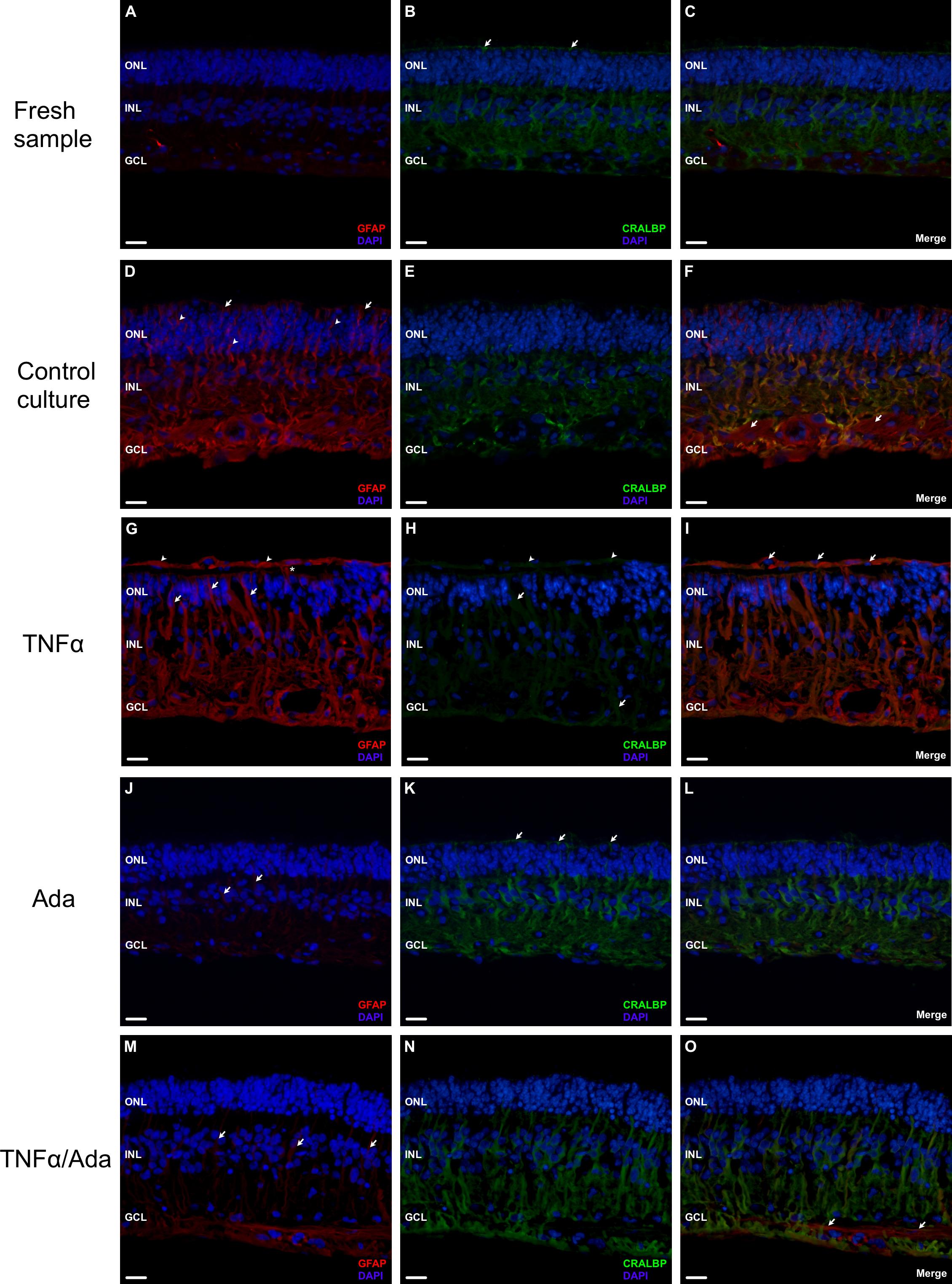
 Figure 1 of
Fernandez-Bueno, Mol Vis 2013; 19:894-903.
Figure 1 of
Fernandez-Bueno, Mol Vis 2013; 19:894-903.