Figure 6. Electromagnetic fields and retinal cells can affect the expression of MMP-2. Expression of MMP-2 in HFSFs were cultured at
ELF-EMFs with RPE supernatant medium or not. The control cells were cultured without ELF-EMFs and RPE supernatant additive.
Then the cells were labeled with antibodies for MMP-2 (green). 4', 6-diamidino-2-phenylindole dyed the nucleus (blue). A-C: PBS was used instead of primary antibody as a negative control. D-F: Control cells were cultured without ELF-EMFs and RPE supernatant additive. G-I: Cells were cultured with ELF-EMFs exposure. MMP-2 showed more marked fluorescent signal than the control cells. J-K: Cells were coexposed to ELF-EMFs and RPE supernatant medium. MMP-2 staining became stronger than exposure to ELF-EMFs only.
Scale bar=20 μm.
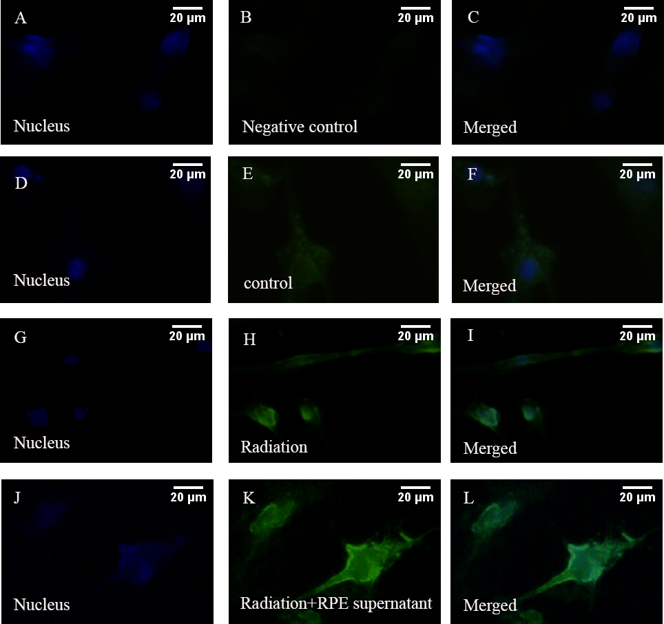
 Figure 6 of
Wang, Mol Vis 2013; 19:885-893.
Figure 6 of
Wang, Mol Vis 2013; 19:885-893.