Figure 5. Electromagnetic fields and retinal cells can affect the expression of COL1A1. The control cells were cultured without ELF-EMFs
and RPE supernatant medium. Then the cells were labeled with antibodies for COL1A1 (green). 4', 6-diamidino-2-phenylindole
dyed the nucleus (blue). A–C: PBS was used instead of primary antibody as a negative control. D–F: Control cells were cultured without ELF-EMFs and RPE supernatant additive. COL1A1 was localized to the cytoplasm. G–I: Cells were cultured after exposure to ELF-EMFs. COL1A1 staining became weak. J–K: Cells were coexposed to ELF-EMFs and RPE supernatant medium. COL1A1 staining much weaker. The scale bar is 20 μm.
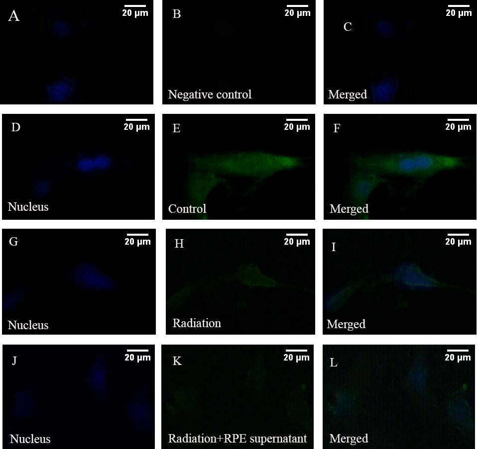
 Figure 5 of
Wang, Mol Vis 2013; 19:885-893.
Figure 5 of
Wang, Mol Vis 2013; 19:885-893.