Figure 3. Clinical images of Case 1. A: A slit-lamp photograph of Case 1 showing a posterior polymorphous dystrophy band lesion (arrows). B: In vivo confocal microscopy in this patient shows undulation of Descemet’s membrane and the endothelial surface, with needle-shaped
hyper-reflectivity at the level of Descemet’s membrane. (Scale bar=100 μm)
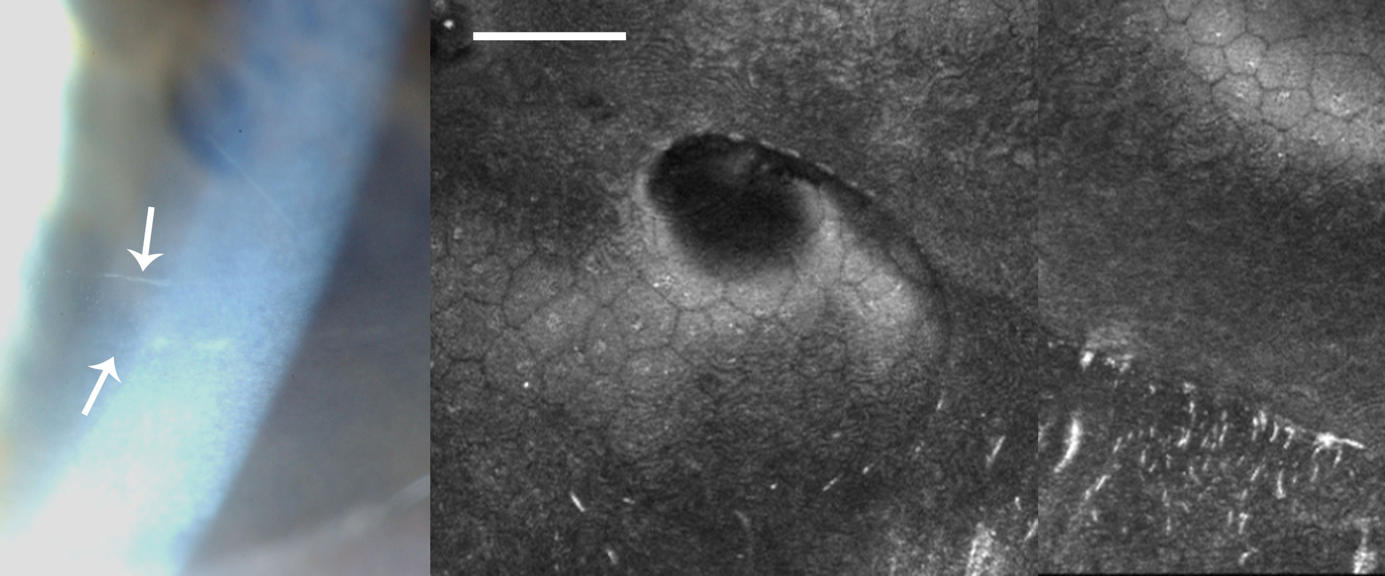
 Figure 3 of
Vincent, Mol Vis 2013; 19:852-860.
Figure 3 of
Vincent, Mol Vis 2013; 19:852-860.