Figure 5. Interleukin 17 Receptor A expression and MIP3α secretion by HCE cells stimulated by interleukin 17 A. A, B: Interleukin 17 Receptor A (IL-17RA) expression was analyzed with immunogold labeling and electron microscopy in HCE cells
either untreated or exposed to interleukin (IL-)17A, respectively. Control unexposed cells expressed IL-17RA in the cytosol.
In IL-17A-exposed cells, immunogold labeled IL-17RA was present in the nucleus in addition to the cytosol. Bar=2 µm. Insets
show higher magnification of gold-labeled antibody. C: After 72 h, there was a significant increase in MIP3α in supernatants from cells exposed to IL-17A (solid bars) compared
to unexposed controls (open bars), indicating an active receptor for IL-17A. Bars represent the mean value of duplicates from
three independent experiments. Values are expressed as mean±SEM, and statistical significance is indicated with an asterisk
(p<0.05).
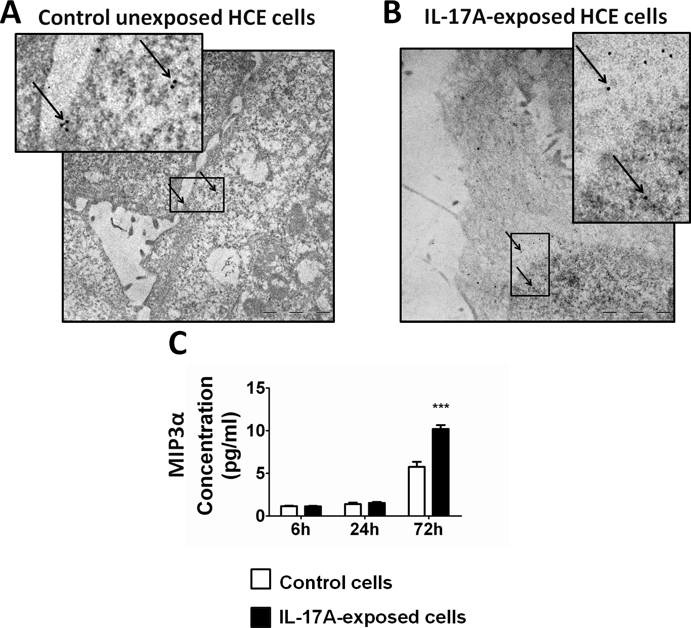
 Figure 5 of
Arranz-Valsero, Mol Vis 2013; 19:85-99.
Figure 5 of
Arranz-Valsero, Mol Vis 2013; 19:85-99.