Figure 3. Interleukin 17A and Interleukin 17 Receptor A expression by HCE cells exposed to Staphylococcus aureus and Pseudomonas aeruginosa supernatants. A: Supernatants, protein, and mRNA from Staphylococcus aureus (SA)- and Pseudomonas aeruginosa (PA)-stimulated HCE cells (solid bars) and from control unstimulated cells (open bars) were collected at different time points.
Bars represent the mean value of duplicates from three independent experiments. Values are expressed as mean±SEM, and statistical
significance, when compared to control unstimulated cells, is indicated with asterisks (*p<0.05). Interleukin (IL-) 17A was
secreted in a time-dependent manner that was not altered under any condition. IL-17A mRNA expression remained constant in
the control unstimulated cells. In stimulated cells, it was significantly decreased at 6 h and tended to be lower at 24 h
and 72 h than in the control cells. IL-17 Receptor A (IL-17RA) protein and mRNA expression did not change significantly with
time. B: Internal control for specific band determination: K562 and HCE protein lysates, and representative western blot images for
each condition: M- Molecular weight markers; 1- SA, 6 h; 2- PA, 6 h; 3- SA, 24 h; 4- PA, 24 h; 5- SA, 72 h; 6- PA, 72 h; 7-
C, 72 h.
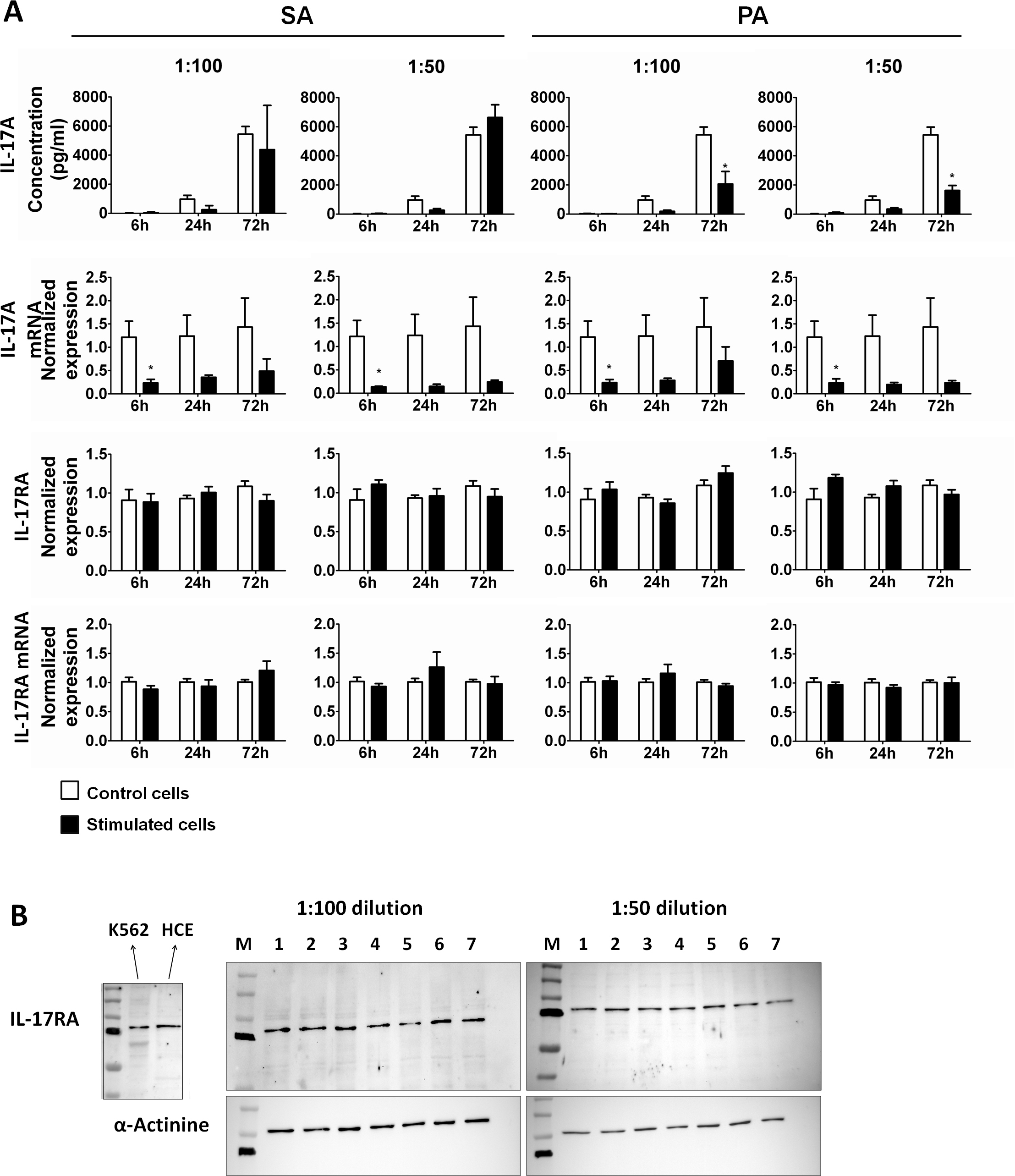
 Figure 3 of
Arranz-Valsero, Mol Vis 2013; 19:85-99.
Figure 3 of
Arranz-Valsero, Mol Vis 2013; 19:85-99.