Figure 2. Interleukin 17 Receptor A expression in human ocular surface tissues. Ocular surface tissues (cornea, limbus, and conjunctiva)
expressed histochemically detectable Interleukin 17 Receptor A (IL-17RA) as seen in these representative micrographs. The
superficial layers were the most intensely stained, and the deeper cells had different patterns depending on the location:
dotted staining in the cornea, staining in the cell-to-cell-contact regions in the limbus, and homogeneous staining in the
conjunctiva. Insets show higher magnification of staining distribution. Bar=40 µm.
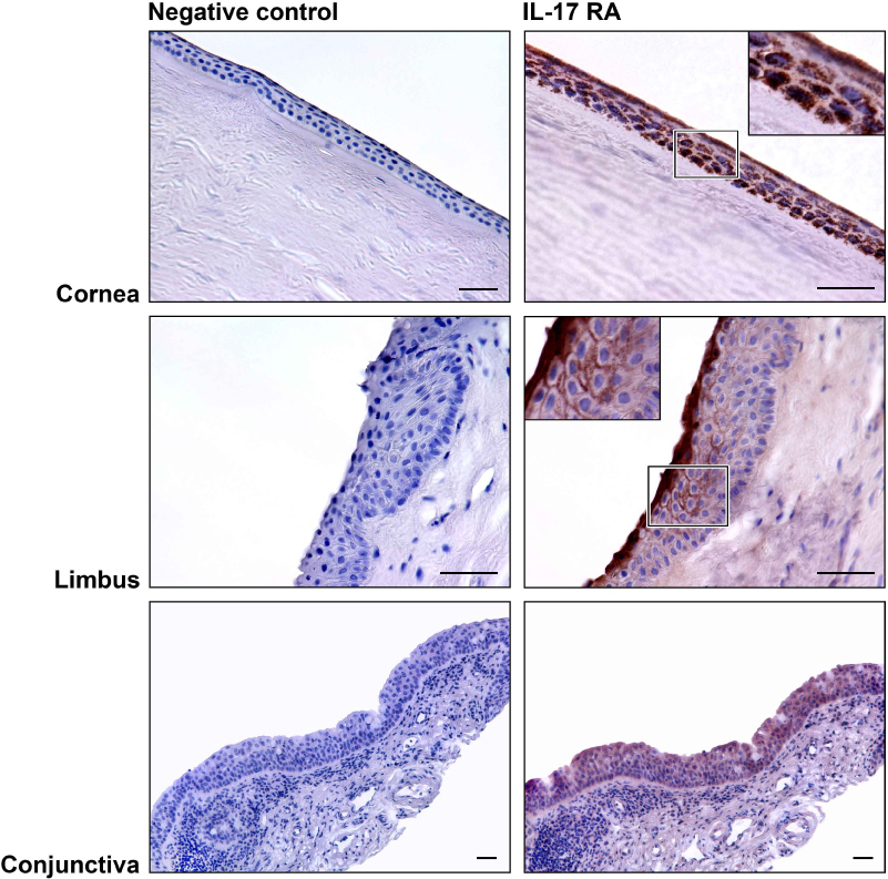
 Figure 2 of
Arranz-Valsero, Mol Vis 2013; 19:85-99.
Figure 2 of
Arranz-Valsero, Mol Vis 2013; 19:85-99.