Figure 4. Mouse ferritin light chain-1 (FTL1) and human ferritin light chain (FTL) transcripts are expressed in the lens. A: Low magnification image (sagittal plane) of the mouse eye (postnatal day 21) shows strong localization of ferritin light
chain-1 (FTL1) transcripts (red punctate staining) to the equatorial epithelium and peripheral cortical fiber cells of the
lens (Co - cornea, CB - ciliary body, and iris, Le - lens, Re - retina). B and C: High-magnification images of FTL1 transcripts expressed in the lens anterior epithelium (B) and equatorial “bow” region (C). Scale bars represent 100 µm. Cell nuclei are stained blue. D: Reverse-transcription polymerase chain reaction (RT–PCR) analysis of lens RNA detects amplicons containing complete coding
sequence for FTL and FTL1. Note the FTL1 amplicon (184 codons) is expected to be slightly larger than that of FTL (176 codons).
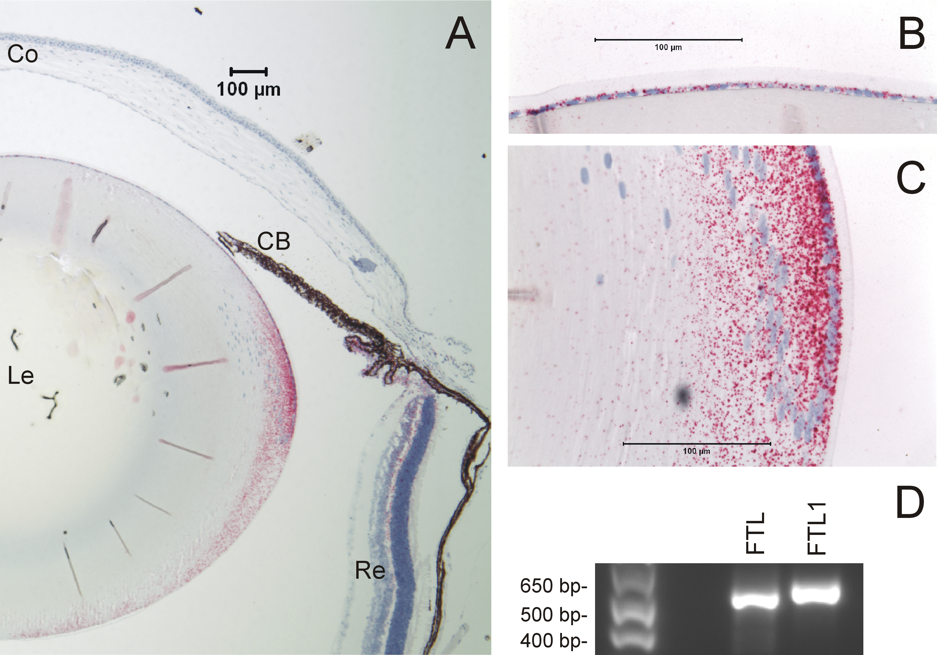
 Figure 4 of
Bennett, Mol Vis 2013; 19:835-844.
Figure 4 of
Bennett, Mol Vis 2013; 19:835-844.