Figure 2. Color fundus photographs of the proband at 43 years of age. Left (A) and right (B) eyes showing typical advanced retinitis pigmentosa changes with bone spicule-like pigment deposits in the posterior pole
and midperiphery along with retinal atrophy, narrowing of the vessels, and waxy optic discs.
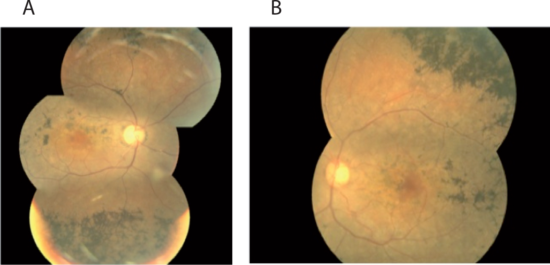
 Figure 2 of
Tiab, Mol Vis 2013; 19:829-834.
Figure 2 of
Tiab, Mol Vis 2013; 19:829-834.